
THE CELL
Introduction
- The cell is the basic unit of an organism.
- All living organisms are made up of cells.
- Some organisms are made up of one cell, while others are multicellular.
- Cells are too small to see with the naked eye.
- They can only be seen with the aid of a microscope.
The Microscope
The microscope is used to magnify objects.
Magnification
- The magnifying power is usually inscribed on the lens.
- To find out how many times a specimen is magnified, multiply the magnifying power of the objective lens by that of the eyepiece lens.
- If the eyepiece magnification lens is x10 and the objective lens is x4, the total magnification is x40.
- Magnification has no units.
- It should always have the multiplication sign, e.g., x40.
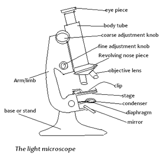
Microscope Parts and Their Functions
| Parts | Function(s) |
|---|---|
| Eyepiece | Has a lens which contributes to the magnification of the object under view. |
| Coarse adjustment knob | Moves the body tube up and down for long distances and brings the image into focus. |
| Fine adjustment knob | Moves the body tube and brings the image into fine focus. |
| Body tube | Holds the eyepiece and the revolving nosepiece. It directs light from objective lenses to the eyepiece lens. |
| Revolving nosepiece | Holds and brings objective lenses into position. |
| Objective lens | Contributes to the magnification of the object. |
| Arm/limb | Used for handling the microscope and tilting it. |
| Stage | Flat platform onto which the slide with the object is placed. |
| Clips | Hold the slide firmly onto the stage. |
| Condenser | Concentrates light onto the object. |
| Diaphragm | Regulates the amount of light passing through the object. |
| Mirror | Reflects light into the condenser. |
| Hinge screw | Fixes the arm to the base and allows for tilting of the arm. |
| Base/stand | Provides support to the microscope. |
To View the Object
- Turn the low power objective lens until it clicks into position.
- Looking through the eyepiece, ensure that enough light is passing through by adjusting the mirror.
- This is indicated by a bright circular area known as the field of view.
- Place the slide containing the specimen on the stage and clip it into position.
- Make sure that the specimen is in the centre of the field of view.
- Using the coarse adjustment knob, bring the low power objective lens to the lowest point.
- Turn the knob gently until the specimen comes into focus.
- If finer details are required, use the fine adjustment knob.
- When using high power objective, always move the fine adjustment knob upwards.
Care of a Microscope
- Great care should be taken when handling it.
- Keep it away from the edge of the bench when using it.
- Always hold it with both hands when moving it in the laboratory.
- Clean the lenses with special lens cleaning paper.
- Make sure that the low power objective clicks in position in line with the eyepiece lens before and after use.
- Store the microscope in a dust-proof place free of moisture.
Cell Structure as Seen Through the Light Microscope


The cell as seen above has the following:
Cell membrane (Plasma membrane):
- This is a thin membrane enclosing cell contents.
- It controls the movement of substances into and out of the cell.
Cytoplasm:
- This is a jelly-like substance in which chemical processes are carried out.
- Scattered all over the cytoplasm are small structures called organelles.
- Like an animal cell, the plant cell has a cell membrane, cytoplasm, and a nucleus.
Vacuole:
- Plant cells have a permanent, central vacuole. It contains cell sap where sugars and salts are stored.
Cell wall:
- This is the outermost boundary of a plant cell.
- It is made of cellulose.
- Between the cells is a middle lamella made of calcium pectate.
Chloroplasts:
- With special staining techniques, it is possible to observe chloroplasts.
- These are structures which contain chlorophyll, the green pigment responsible for trapping light for photosynthesis.
The Electron Microscope (EM)
- Capable of magnifying up to 500,000 times.
- The specimen is mounted in a vacuum chamber through which an electron beam is directed.
- The image is projected onto a photographic plate.
- The major disadvantage of the electron microscope is that it cannot be used to observe living objects.
- However, it provides a higher magnification and resolution (ability to see close points as separate) than the light microscope so that specimens can be observed in more detail.
Cell Structure as Seen Through Electron Microscope


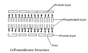
The Plasma Membrane
- Under the electron microscope, the plasma membrane is seen as a double layer.
- This consists of a lipid layer sandwiched between two protein layers.
- This arrangement is known as the unit membrane and shows two lipid layers with proteins within.
- Substances are transported across the membrane by active transport and diffusion.
The Endoplasmic Reticulum (ER)
- This is a network of tubular structures extending throughout the cytoplasm of the cell.
- It serves as a network of pathways through which materials are transported from one part of the cell to another.
- An ER encrusted with ribosomes is referred to as rough endoplasmic reticulum.
- An ER that lacks ribosomes is referred to as smooth endoplasmic reticulum.
- The rough endoplasmic reticulum transports proteins while the smooth endoplasmic reticulum transports lipids.
The Ribosomes
- These are small spherical structures attached to the ER.
- They consist of protein and ribonucleic acid (RNA).
- They act as sites for the synthesis of proteins.
Golgi Bodies
- Golgi bodies are thin, plate-like sacs arranged in stacks and distributed randomly in the cytoplasm.
- Their function is packaging and transportation of glycoproteins.
- They also produce lysosomes.
Mitochondria
- Each mitochondrion is a rod-shaped organelle.
- Made up of a smooth outer membrane and a folded inner membrane.
- The foldings of the inner membrane are called cristae.
- They increase the surface area for respiration.
- The inner compartment is called the matrix.
- Mitochondria are the sites of cellular respiration, where energy is produced.
Lysosomes
- These are vesicles containing hydrolytic enzymes.
- They are involved in the breakdown of micro-organisms, foreign macromolecules, and damaged or worn-out cells and organelles.
The Nucleus
- The nucleus is surrounded by a nuclear membrane which is a unit membrane.
- The nuclear membrane has pores through which materials can move to the surrounding cytoplasm.
- The nucleus contains proteins and nucleic acids deoxyribonucleic acid (DNA) and RNA.
- The chromosomes are found in the nucleus.
- They are the carriers of the genetic information of the cell.
- The nucleolus is also located in the nucleus but it is only visible during the non-dividing phase of the cell.
The Chloroplasts
- These are found only in photosynthetic cells.
- Each chloroplast consists of an outer unit membrane enclosing a series of interconnected membranes called lamellae.
- At various points along their length, the lamellae form stacks of disc-like structures called grana.
- The lamellae are embedded in a granular material called the stroma.
- The chloroplasts are sites of photosynthesis.
- The light reaction takes place in the lamellae while the dark reactions take place in the stroma.
Comparison Between Animal Cell and Plant Cell
| Plant Cell | Animal Cell | |
|---|---|---|
| Has a cell wall and a cell membrane. | Has cell membrane only. | |
| Nucleus at periphery. | Nucleus at the center. | |
| Have chloroplasts. | Have no chloroplasts. | |
| Has a large central vacuole. | Has no vacuoles; they are small and scattered. | |
| Are usually large. | Are usually small. | |
| Are regular in shape. | Irregular in shape. | |
| Has no centriole. | Has centrioles. | |
| Stores starch, oils, and protein. | Stores glycogen and fats. |
Cell Specialisation
Cells are specialised to perform different functions in both plants and animals.
Example:
- Palisade cells have many chloroplasts for photosynthesis.
- Root hair cells are long and thin to absorb water from the soil.
- Red blood cells have haemoglobin which transports oxygen.
- Sperm cells have a tail to swim to the egg.
- In multicellular organisms, cells that perform the same function are grouped together to form a tissue.
- Each tissue is therefore made up of cells that are specialised to carry out a particular function.
Animal Tissues – Examples of Animal Tissues
| Type of Tissue | Functions | Characteristics |
|---|---|---|
| Epithelial Tissue | Covering, allowing movement of materials. | |
| Squamous epithelium | Covering of internal organs, lining for body cavity. | Thin flat cells. |
| Columnar epithelium | Secretion, absorption e.g., in the alimentary canal. | Cells that are longer than they are wide. |
| Stratified epithelium | Covering surfaces, protection e.g., the skin. | Several layers of epithelial cells (either squamous, cuboidal, or columnar). |
| Cuboidal epithelium | Absorption e.g., in the kidney tubules. | Cube-like cells. |
| Muscular Tissue | Contraction, bringing about movement of body parts. | Consists of units called myofibrils. |
| Striated (skeletal or voluntary muscle) | Contract and allow movement. | Are multinucleated; have transverse striations; controlled by voluntary nervous system. |
| Smooth (visceral or involuntary muscle) | Cover internal organs; allow movement e.g., peristalsis. | Are spindle-shaped, mononucleated; controlled by involuntary nervous system. |
| Cardiac muscle | Cause contraction of the heart. | Contract rhythmically; are myogenic (ability to contract is within). |
| Supporting Tissue | Support the body, provide a rigid framework, protect soft tissue. | Cells that produce hard materials. |
| Cartilage | ||
| Bone | ||
| Blood | Transport of materials, protection against disease. | A complex tissue consisting of three types of cells suspended in a fluid medium (Plasma). |
| Nerve Tissue | Receive stimuli and transmit impulses; coordinate body activities. | Consists of cells called neurones which are interconnected through axons to enable transmission of impulses. |
Plant Tissues
Example of Plant Tissues
| Type of Tissue | Functions | Characteristics |
|---|---|---|
| Meristematic | Undergo division and cause growth, e.g., increase in length and girth. | Small thin-walled cells, contain a lot of cytoplasm; found mostly at the tip of shoots and roots. |
| Parenchyma | Photosynthesis, gaseous exchange, support, storage. | Thin-walled cells; vary in shape and size; many intercellular spaces. |
| Collenchyma | Strengthening. | Thickened walls; no intercellular spaces; found in cortex of stems. |
| Sclerenchyma | Strengthening. | Vary in shape; thick cell walls; are usually dead. |
| Vascular | Transport materials. | Tubular vessels and tracheids. |
| Xylem | Transport of water and mineral salts. | Joined end to end. |
| Phloem | Transport of organic materials (manufactured food). | Sieve elements joined to each other through sieve pores. |
Organs
- An organ is made up of different tissues.
- Examples include the heart, lungs, kidneys, and brain in animals, and roots, stems, and leaves in plants.
Organ Systems
- Organs which work together form an organ system.
- Examples include digestive, excretory, nervous, and circulatory systems in animals, and transport and support systems in plants.
Organism
- Different organ systems form an organism.
Practical Activities
Observation and Identification of Parts of a Light Microscope and Their Functions:
- A light microscope is provided.
- Various parts are identified and observed.
- Drawing and labelling of the microscope is done.
- Functions of the parts of the microscope are stated.
- Calculations of total magnification done using the formula:
- Eyepiece lens magnification x objective lens magnification.
Preparation and Observation of Temporary Slides of Plant Cells
- A piece of epidermis is made from the fleshy leaf of an onion bulb. It is placed on a microscope slide and a drop of water added.
- A drop of iodine is added and a cover slip placed on top.
- Observations are made under low and medium power objectives.
- The cell wall and nucleus stain darker than other parts.
- A labelled drawing is made.
- The following are noted: nucleus, cell wall, cytoplasm, and cell membrane.
Observation of Permanent Slides of Animal Cells
- Permanent slides of animal cells are obtained, e.g., of cheek cells, nerve cells, and muscle cells.
- The slide is mounted on the microscope and observations made under low power and medium power objectives.
- Labelled drawings of the cells are made.
- A comparison between plant and animal cells is made.
Observation and Estimation of Cell Size and Calculation of Magnification of Plant Cells
- Using the low power objective, a transparent ruler is placed on the stage of the microscope.
- An estimation of the diameter of the field of view is made in millimeters.
- This is converted into micrometres (1 mm = 1000 µm).
- A prepared slide of onion epidermal cells is mounted.
- The cells across the centre of the field of view are counted from left to right and top to bottom.
- The diameter of the field of view is divided by the number of cells lying lengthwise to give an estimate of the length and width of each cell.


1 Comment