
Significance of Meiosis
Meiosis ensures a constant chromosome number for all species that reproduce sexually. During gamete formation, the chromosome number is halved and then restored at fertilization, maintaining genetic stability across generations.
Additionally, meiosis provides opportunities for new gene combinations through the formation of chiasmata, which is a key mechanism for genetic variation and evolution.
Difference Between Meiosis and Mitosis
| Stage | Mitosis | Meiosis |
|---|---|---|
| Prophase |
|
|
| Metaphase | Chromatid pairs line up on the equator of the spindle; centromeres line up on the equator. |
|
| Anaphase |
|
|
| Telophase | Both homologous chromosomes are in each daughter cell | Only one of each pair of homologous chromosomes is in each daughter cell |
| Occurrence | Occurs in the formation of somatic cells | Occurs in the formation of gametes and spores |
Stages of Sexual Reproduction
Sexual reproduction involves the following stages:
- Gametogenesis
- Copulation
- Fertilization
- Cleavage
- Implantation
- Pregnancy
- Parturition (birth)
- Parental care
Gametogenesis
Definition: Gametogenesis is the general process of gamete formation in both male and female organisms reproducing sexually.
- Meiosis is the process by which gametes are formed; it is also called gametogenesis, literally meaning ‘creation of gametes’.
- In males, meiosis transforms spermatogonia into primary spermatocytes, then secondary spermatocytes, spermatids, and finally spermatozoa; this process is called spermatogenesis.
Definition: Oogenesis is the process of meiosis in females, transforming oogonia into primary oocytes, secondary oocytes, and ultimately an ovum (egg cell).
- Primordial germ cells populate the gonads and proliferate into sperm in testes or ova in ovaries.
Spermatogenesis
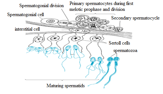
- In the male testis, seminiferous tubules contain diploid cells called spermatogonia that develop into mature spermatozoa, the male gametes.
- Spermatogonia multiply by mitosis to maintain their population and produce spermatocytes.
- Specialized spermatogonia are located around the periphery of the seminiferous tubules.
Spermatogonia are diploid cells set aside early in embryonic development. They divide by mitosis to generate more spermatogonia or enter meiosis to produce spermatids, which differentiate into mature sperm cells.
- Spermatogonia destined for meiosis differentiate into primary spermatocytes, which undergo two successive meiotic divisions.
- After meiosis I, secondary spermatocytes are formed; these undergo meiosis II to become spermatids, each with a unique set of 23 chromosomes, which mature into four spermatozoa.
The Seminiferous Tubules Contain Two Types of Cells:
- Germ cells: Undergo meiosis to form spermatozoa.
- Sertoli Cells: Nurse cells that provide nourishment to germ cells.
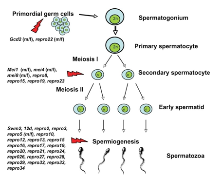
Fig: The stages of spermatozoa formation.
- Spermatids undergo transformation to become spermatozoa.
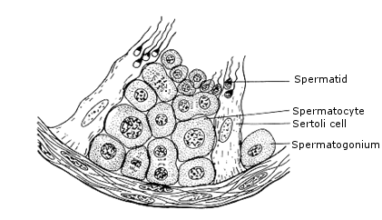
Fig: Diagram showing the structure of part of the wall of seminiferous tubule.
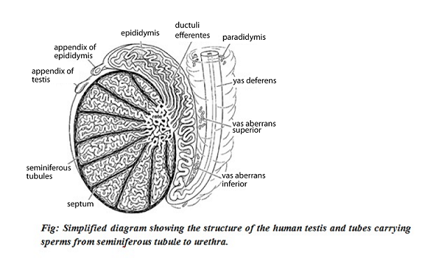
Spermatogenesis
- Occurs in seminiferous tubules.
- Stored in epididymis.
Process:
- Diploid spermatogonia divide by mitosis from germinal epithelium.
- Some grow into diploid primary spermatocytes.
- Primary spermatocytes undergo first meiotic division to form two haploid secondary spermatocytes.
- Secondary spermatocytes undergo second meiotic division to form haploid spermatids.
- Spermatids mature into spermatozoa.
- Sertoli cells provide nutrition and protect germ cells from the immune system.
From the figure:
The interior of the testis is the site of spermatogenesis within seminiferous tubules. Spermatogonia develop into sperm through spermatocyte and spermatid stages. Each sperm has a long tail and a head containing a haploid nucleus.

Mechanism of Spermatogenesis
Spermatogenesis is divided into four phases:
- Multiplication phase
- Growth phase
- Maturation phase
- Metamorphosis
1. Multiplication Phase
- Also called spermatocytogenesis.
- Sperm mother cells in the germinal epithelium divide repeatedly by mitosis to form many diploid spermatogonia.
Some spermatogonia move toward the lumen of seminiferous tubules and enter the growth phase as primary spermatocytes, which are diploid with (44 + XY) chromosomes.
Some spermatogonia remain as stem cells, continuing to divide and produce primary spermatocytes.
2. Growth Phase
The spermatocyte and its nucleus enlarge in preparation for meiotic division.
3. Maturation Phase
Each diploid primary spermatocyte undergoes meiosis I (reduction division), producing two haploid secondary spermatocytes with (22 + X) or (22 + Y) chromosomes.
Secondary spermatocytes undergo meiosis II (equational division), forming four haploid spermatids.
4. Metamorphosis
Spermatids are typical animal cells with organelles but cannot function as gametes. They undergo changes to become motile spermatozoa, increasing motility through:
- Nucleus shrinks and DNA condenses.
- Acrosome forms from the Golgi complex.
- Axial filament of the tail forms from the distal centriole.
- Mitochondrial ring forms around distal centrioles.
- Cytoplasm is mostly lost; remaining cytoplasm forms a sheath called the manchette.
- Developing sperms have their heads embedded in Sertoli cells, which provide nutrition via glycogen stores that diminish as spermatids mature.
Note: Although direct evidence is lacking, failure to produce normal Sertoli cells is linked to male sterility.
Cellular Events in Human Spermatogenesis
Sertoli cells support developing gametes by:
- Maintaining the environment necessary for development via the blood-testis barrier.
- Secreting substances that initiate meiosis.
- Producing supporting testicular fluid.
- Secreting androgen-binding protein (ABP) to concentrate testosterone near developing gametes.
Testosterone is produced by Leydig cells adjacent to seminiferous tubules.
- Secreting hormones like inhibin that regulate pituitary control of spermatogenesis.
- Phagocytizing residual bodies from spermiogenesis.
- Releasing anti-Mullerian hormone (AMH) to prevent formation of the Mullerian duct.
Note: Seminiferous epithelium is sensitive to elevated temperatures and functions optimally at 2–8°C below body temperature, which is maintained by the scrotum through blood flow regulation and muscle positioning.
- Factors such as vitamin deficiencies, anabolic steroids, metals, X-rays, dioxin, alcohol, and infections can adversely affect spermatogenesis.
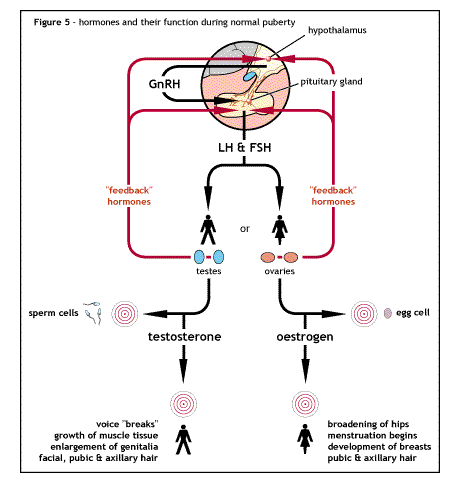
- Hormonal control involves the hypothalamus, pituitary gland, and Leydig cells, with key hormones being luteinizing hormone (LH), follicle-stimulating hormone (FSH), and testosterone.
- LH stimulates testosterone secretion by Leydig cells, which acts on Sertoli cells to increase their responsiveness to FSH and inhibits LH secretion via feedback.
- FSH promotes maturation of spermatic epithelium by acting on Sertoli cells.
- ABP is produced by Sertoli cells to bind and transport androgens.
- FSH is necessary for ABP production and development of the blood-testis barrier.
Once Sertoli function is established, testosterone alone can maintain spermatogenesis, but FSH increases sperm yield by preventing atresia of differentiating spermatogonia.
FSH levels are influenced by environment, sexual activity, and inhibin.
- Androgens produced by Leydig cells influence developing germ cells, assisted by ABP produced by Sertoli cells.
Testosterone induces and maintains spermatogenesis via Sertoli or germ cells.
- Other testicular hormones include:
i) Estradiol: Estrogen receptors in testicular cells suggest a role in testicular function regulation.
ii) Inhibin (Inh-b): Produced by Sertoli cells, it controls FSH secretion via negative feedback; low levels indicate spermatogenesis disorders.
iii) Anti-Mullerian hormone: Secreted by Sertoli cells, influenced by testosterone, FSH, and spermatocytes; prevents Mullerian duct formation.
Summary: Mechanism of Hormonal Control of Spermatogenesis
- The hypothalamus secretes gonadotrophin releasing hormone (GnRH) to stimulate the anterior pituitary gland.
- The pituitary releases gonadotrophins: FSH and LH.
- FSH stimulates Sertoli cells to support sperm development.
- LH stimulates Leydig cells to produce testosterone.
- Testosterone promotes germinal epithelial growth and works with FSH on Sertoli cells.
Negative feedback reduces GnRH, LH, and FSH secretion when testosterone levels rise. Inhibin released during high spermatogenesis reduces FSH secretion.
The Role of Cyclic AMP
FSH and LH act by releasing cyclic AMP (cAMP) inside target cells. cAMP acts as a second messenger, stimulating enzyme synthesis in the nucleus. For example, LH stimulates enzymes for testosterone synthesis from cholesterol.

Structure of Mature Human Spermatozoa
A spermatozoan is divided into three parts:
I) Head Piece
- Contains the nucleus and a small amount of cytoplasm.
- The acrosome at the tip contains hydrolytic enzymes like proteases and hyaluronidases to digest the egg membrane.
II) Middle Piece
Contains mitochondria that provide energy for sperm motility. The head and middle piece together form the principal piece.
III) Tail Piece
- Consists of the flagellum made of axial filaments continuing from the middle piece.
- Propels the sperm toward the egg and orients it for proper binding.
- The end piece is a hair-like extension at the tail’s end.

Role of Spermatozoan
- To deliver the paternal genetic material into the egg so that the zygote inherits a combination of maternal and paternal genes.
Hormonal Control of Sperm Production
- Low testosterone levels stimulate the hypothalamus to secrete GnRH.
- GnRH stimulates the pituitary to release LH and FSH.
- LH triggers Leydig cells to produce testosterone.
- FSH stimulates Sertoli cells to support sperm formation.
- Rising testosterone levels inhibit GnRH, LH, and FSH secretion.
- Testosterone also causes Sertoli cells to release inhibin, which inhibits FSH production.
When testosterone drops, the cycle restarts.

Adaptations of the Spermatozoa
Spermatozoa are adapted to their function by:
- Having an acrosome with enzymes to digest the egg membrane.
- Containing numerous mitochondria to provide energy for movement.
- Having a flagellum to propel the sperm toward the egg.
- Being able to sense chemical attractants secreted by the egg.
- Recognizing and binding to receptor sites on the egg surface.
- Having a light nucleus and streamlined head to move quickly.
Oogenesis
- Begins soon after fertilization as primordial germ cells migrate to the gonads and proliferate mitotically.
- Germ cells multiply from a few thousand to nearly 7 million.
- Oocytes enter meiosis and arrest in prophase I until puberty.
- At puberty, 4 to 10 follicles develop, but only 1–2 oocytes are released.
- Each oocyte is surrounded by zona pellucida and follicle cells.
- First meiotic division produces a secondary oocyte and polar body.
- Secondary oocyte begins second meiotic division but arrests at metaphase II until fertilization.
Summary: Oogenesis
At Birth:
- Diploid oogonia divide by mitosis.
- Oogonia undergo meiosis I to form primary oocytes arrested at prophase I.
- Primary oocytes remain in follicles.
At Puberty:
- Primary oocyte completes meiosis I, forming secondary oocyte and polar body.
- Secondary oocyte undergoes meiosis II and arrests at metaphase II.
- If fertilized, secondary oocyte completes meiosis II to form ovum and polar bodies.
- Ovum formation involves increased cell volume and organelle acquisition to support fertilization and early embryo development.
- Most oogonia are destroyed before birth, leaving a finite number of oocytes at birth.
Unlike spermatogenesis, oogenesis produces one large ovum and three small polar bodies due to unequal cytoplasmic division.
Egg cytoplasm contains abundant RNA types that direct early embryonic protein synthesis and development.
Development of Germ Cells in the Ovary
Primordial germ cells migrate into the gonadal ridge, proliferate, and are enveloped by coelomic epithelial cells forming germinal cords.
Processes include:
- Proliferation from 15th week to 7th month of gestation.
- Primary germ cells arise via mitosis of oogonia connected by cellular bridges for synchronous meiosis onset.
- Designation changes to primary oocytes as meiosis begins in prophase I around 12th week.
- Primary oocytes arrest in diplotene stage of prophase I until puberty.
This meiotic resting phase, called dictyotene, lasts until puberty when oocytes resume meiosis monthly.
- Only a few oocytes complete meiosis and ovulate; others become atretic.
- Primary oocytes may remain arrested until menopause.
- Two structures develop asynchronously: primary oocytes and follicular epithelium.
- Primary oocytes develop further only after puberty with hormonal stimulation.
- Follicular epithelium develops through follicle stages while oocytes remain primary.
The developmental sequence of female germ cells is shown below:
Oocyte maturation resumes only days before ovulation.
- Follicle development proceeds through various stages.
- Many follicles undergo atresia and do not reach the tertiary stage.
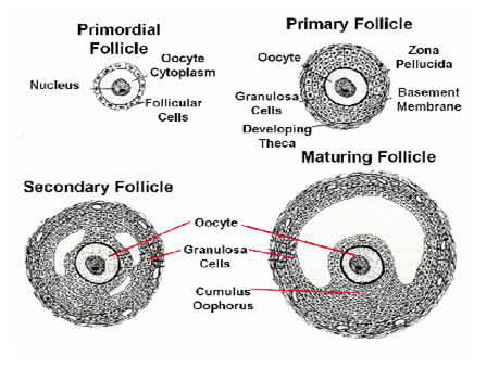
Primary Oocyte
- Oocyte maturation depends on follicle cell development.
- The dominant follicle matures into the Graafian follicle.
- Oocyte ingests substances and yolk supplied by granulosa cells via cytoplasmic processes through the zona pellucida.
- The oocyte nucleus matures before the LH peak.
The oocyte remains arrested in the prolonged prophase (dictyotene) of meiosis I since fetal life.
- Maturation prepares the nucleus for meiosis completion triggered by the LH peak.
LH triggers maturation steps leading to ovulation.
In the Oocyte:
- Completion of meiosis I with first polar body ejection.
- Start of meiosis II with arrest at metaphase.
- Cytoplasmic maturation preparing for fertilization.
In the Follicle:
- Granulosa cells retract processes from the oocyte surface.
- Perivitelline space forms between oocyte and zona pellucida, housing the first polar body.
- Granulosa cells loosen and proliferate near the cumulus oophorus.
- Progesterone concentration increases in follicle fluid.
Termination of the First Meiosis
- Spindle apparatus forms and orients radially on the cell surface.
- First polar body forms at spindle attachment site.
Granulosa cell retraction leads to perivitelline space formation where the first polar body is ejected, marking meiosis I completion.
- Primary oocyte becomes secondary oocyte.
The Secondary Oocyte
- LH stimulates granulosa cells to lose processes and multiply.
- Granulosa cells produce progesterone released into follicle fluid.
- Secondary oocyte contains a haploid (duplicated) chromosome set after meiosis I.
Role of Progesterone in Follicle Fluid
- Stimulates further oocyte maturation.
- During ovulation, guides sperm by forming a chemical gradient in the fallopian tubes.
Follicle About to Rupture
- Granulosa cells secrete extracellular matrix rich in hyaluronic acid into follicle fluid.
The cumulus oophorus loosens, freeing the oocyte and surrounding granulosa cells (corona radiata) from the follicular wall.
- Oocyte completes maturation steps triggered by LH peak.
- Preparations for fertilization occur in cytoplasm.
- Spindle apparatus forms for meiosis II, arrested at metaphase.
Note: Meiosis II is arrested until sperm penetration.
- Ovulation occurs about 38 hours after LH peak.
Adaptation of the Egg Cell
- Microvilli for nutrient absorption from follicular cells.
Follicular cells surround the ovum but are not part of the egg cell.
- Stored food (yolk) for zygote and embryo development.


Fig: The stage of oogenesis
- Cortical granules act as vesicles to prevent polyspermy during fertilization.
This occurs by:
- Cortical granules fuse with the membrane after sperm entry, releasing chemicals that harden the membrane, forming a fertilization membrane to block additional sperm.
- Destruction of sperm receptor sites on the ovum immediately after sperm entry.
- Receptor sites for sperm binding during fertilization.
- Production of chemicals attracting sperm.
Differences Between Spermatozoa and Egg Cell
| Spermatozoa | Egg Cell | |
|---|---|---|
| Small in size. | Larger than sperm. | |
| Smaller nucleus. | Large nucleus. | |
| Very little cytoplasm. | Large amount of cytoplasm. | |
| No food reserves. | Stores large amounts of food. | |
| Has acrosome. | No acrosome. | |
| No cortical granules. | Has cortical granules. | |
| Has head, middle piece, principal piece, and end piece. | No such division. | |
| No microvilli. | Has microvilli. | |
| Single layered. | Multi-layered. | |
| Flagellated and motile. | Lacks flagellum and non-motile. | |
| Numerous mitochondria. | Few mitochondria. |
Difference Between Oogenesis and Spermatogenesis
| Spermatogenesis | Oogenesis | |
|---|---|---|
| Differentiation follows meiotic division; cells formed only until meiosis ends. | Egg grows primarily during extended prophase I; secondary oocyte is mature. | |
| Occurs in male gonads (testis). | Occurs in female gonads (ovaries). | |
| Four sperms produced from one spermatogonium. | One ovum produced from one oogonium. | |
| Spermatocyte divides into four equal cells, all becoming spermatozoa. | Oocyte divides unequally producing one large ovum and three small polar bodies. | |
| Spermatozoa produced in large numbers. | Ova produced in limited numbers. | |
| Spermatozoa are minute, yolkless, and motile. | Ova are larger, often yolky, and non-motile. | |
| Continuous production from puberty to old age; quality and quantity fluctuate. | Oocytes generated before birth are used up; numbers decline until menopause. | |
| No meiotic division or germ cell production during fetal period. | Meiosis begins during fetal period and arrests in dictyotene stage. |
Fertilization
Fertilization is the fusion of the male and female gamete nuclei to form a diploid zygote nucleus.
- This occurs in the fallopian tube after sperm undergo capacitation.
Capacitation
Capacitation activates sperm before fertilization, taking about 7 hours, involving:
- Removal of glycoprotein and plasma protein layers from sperm surface by uterine enzymes.
Cholesterol is also removed, increasing membrane permeability to Ca2+ ions.
Calcium ions:
- Increase sperm flagellum beating.
- Promote acrosomal reaction.
Mechanism of Fertilization
Fertilization involves two chemical reactions:
- Acrosomal reactions
- Cortical reactions
Steps:
- Sperm migrates through follicle cell coat and binds to zona pellucida receptors.
- Acrosomal reaction releases enzymes (proteases and hyaluronidase) digesting zona pellucida.
- Sperm reaches egg membrane and binds to receptors, inducing Na+ influx and membrane depolarization (first polyspermy block).
- Plasma membranes fuse, allowing sperm nucleus entry.
- Sperm-egg fusion causes Ca2+ influx, triggering cortical reaction.
Cortical reaction causes zona pellucida hardening and fertilization membrane formation (second polyspermy block).
- Secondary oocyte completes meiosis II, forming ovum. Male and female pronuclei fuse to form zygote.
Note: Unfertilized secondary oocytes degenerate after ovulation.
Post-Fertilization Changes in the Egg
- Zygote prepares for cleavage and embryo formation.
- Oxygen consumption increases.
- Metabolic rate increases, including amino acid uptake and membrane permeability changes.
- Protein synthesis begins.
Significance of Fertilization
- Restores diploid chromosome number.
- Establishes egg polarity and new genetic constitution.
- Activates egg for cleavage.
- Introduces genetic variation through combining parental traits.
- Increases metabolic activity and protein synthesis.
The Concept of Sterility
Sterility: Failure of a mature mammal to fertilize or be fertilized.
Causes of Infertility
| Female Infertility | Male Sterility/Infertility |
|---|---|
| Failure to ovulate due to hormonal causes. | Absence of sperm due to blockage between testes and seminal vesicles. |
| Uterus damage preventing pregnancy maintenance (miscarriage). | Low sperm count. |
| Oviduct damage causing blockage. | Production of abnormal sperm. |
| Cervix damage reducing cervical mucus production. | Autoimmunity attacking sperm, reducing count. |
| Antibodies to sperm. | Impotence. |
Impotence
Failure of the penis to erect, which can be:
- Temporary, caused by depression, fear, or psychological factors.
- Permanent, due to genetic, disease, or hormonal issues.
Copulation ensures sperm transfer from male to female reproductive organs for fertilization.
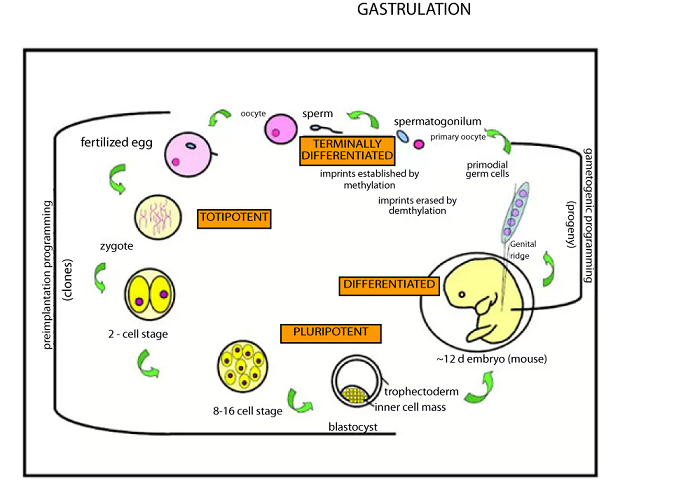
Development of the Zygote and Embryo
Includes five stages:
- Cleavage
- Blastulation
- Gastrulation
- Neurulation
- Organogenesis
Cleavage
- Two hours after fertilization, zygote divides mitotically into two cells (blastomeres).
- Second cleavage forms four blastomeres.
- Cleavage becomes irregular, forming a morula.
- Cleavage does not increase morula size as cells remain in zona pellucida.
- Process lasts about 72 hours.
Cleavage increases surface area to volume ratio, enhancing nutrient uptake, waste removal, and embryonic tissue formation.
Blastulation
Transformation of morula into blastula or blastocyst.
- As zygote moves through oviduct, zona pellucida is shed in uterus.
- Trophoblast forms outer cell layer.
- Inner cell mass forms at one end, creating blastocoel cavity.
Implantation
Blastocyst embeds into uterine wall.
- Trophoblast secretes enzymes digesting endometrium for embedding.
Trophoblast develops villi (chorion villi) for nutrient absorption and secretes human chorionic gonadotrophin (HCG), which:
- Maintains corpus luteum secretion of estrogen and progesterone.
- Inhibits menstruation.
- Forms basis of pregnancy tests.
- Continues uterine wall penetration until fully embedded.
Gastrulation
Blastula transforms into gastrula with germ layers.
- Cells invaginate forming blastopore.
- Germ layers form: ectoderm, mesoderm, endoderm.
Note: Gastrulation is crucial for placenta development and cell fate determination.
Blastopore becomes anus; archenteron forms digestive tract.
Ectoderm: Nervous system, skin, sense organs.
Mesoderm: Bones, muscles, blood, reproductive system.
Endoderm: Digestive and respiratory systems, glands.
Extra Embryonic Membranes and Their Roles
After implantation, four membranes develop outside the embryo:
- Chorion
- Amnion
- Allantois
- Yolk sac
I. Chorion
Outermost membrane from trophoblast cells with villi forming part of placenta.
- Forms placenta parts.
- Absorbs nutrients from mother.
- Protects fetus.
II. Amnion
Innermost membrane lining amniotic cavity filled with fluid cushioning embryo.
III. Allantois
Outgrowth from embryonic gut; fuses with chorion forming placenta; develops into umbilical cord.
IV. Yolk Sac
No obvious function in humans; important in reptiles and birds for nutrient transfer.
Placenta
- Forms when chorionic villi penetrate endometrium.
- Villi surrounded by maternal blood pools (placental sinuses).
- Links fetus and mother; has fetal and maternal sides.
- Exchanges materials by diffusion without blood mixing.
Why Maternal and Fetal Blood Do Not Mix
- Maternal blood pressure is higher and could damage fetal tissues.
- Mixing could trigger maternal immune response against fetus.
- Fetal cells are genetically different due to paternal genes.
Hormones Involved
- Progesterone
- Estrogen
- Human chorionic gonadotrophic hormone
Roles of Placenta
- Allows material exchange without blood mixing.
- Passes oxygen, water, glucose, and nutrients to fetus.
- Removes carbon dioxide, urea, and wastes from fetus.
- Transfers antibodies for passive immunity.
- Protects fetus from some pathogens and toxins.
- Prevents passage of certain hormones and chemicals like alcohol.
Twins Puzzle and Multiple Birth
Multiple Birth and Their Causes
Multiple births occur when more than one baby is born from the same pregnancy.
- Common in mammals like cats, rabbits, dogs, and pigs due to multiple ovulations.
Humans usually have single births.
Causes of Multiple Birth
- More than one secondary oocyte released and fertilized.
- Zygote splits into multiple embryos after fertilization.
Twins
Definition: Two or more babies born from the same pregnancy.
Types of Twins
- Identical Twins
- From one zygote splitting into two or more embryos.
- Share the same placenta, chorion, and amnion.
- Have identical genetic makeup and same sex.
- Rarely, incomplete separation leads to conjoined (Siamese) twins.
Fraternal/Non-identical Twins
- From two different zygotes fertilized by different sperm.
- Each develops in its own placenta and membranes.
- Genetically different and may be different sexes.
Differences Between Identical and Non-Identical Twins
| Identical Twins | Non-Identical Twins | |
|---|---|---|
| Result from one zygote. | Result from two different zygotes. | |
| Share the same placenta. | Each has its own placenta. | |
| Enclosed in the same membrane. | Each has its own membrane. | |
| Same genetic makeup. | Genetically different. | |
| Same sex. | May be different sexes. |
Birth (Parturition)
Birth is the expulsion of the fully developed fetus from the mother’s womb after gestation.
The Process of Birth/Labour
Labour has three stages, longer in first pregnancies:
First Stage
Labour pains begin. The fetal hypothalamus releases ACTRF, stimulating fetal pituitary to release ACTH, which triggers fetal adrenal glands to release corticosteroids. These enter maternal circulation and:
- Increase uterine prostaglandins.
- Decrease progesterone.
- Oxytocin release from maternal pituitary is allowed.
- Myometrium contractions are stimulated by prostaglandins and oxytocin.
Cervix dilates under relaxin influence; amnion and chorion rupture releasing amniotic fluid (“breaking of water”).
Contractions force baby through birth canal; fetal head engages pelvis and cervix.
First stage ends when fetal head diameter equals cervix diameter.
Second Stage
Baby is delivered.
- Umbilical cord is ligated and cut to separate baby from mother.
Third Stage
Delivery of placenta and membranes (afterbirth) occurs due to uterine contractions.
Afterbirth must be expelled promptly to prevent infection.
Parental Care
Includes all activities parents perform for offspring growth and upbringing.
Aspects of Parental Care
- Nutrition (first 3 months): Baby is fed nutritious food, primarily breast milk containing essential nutrients and antibodies (colostrum).
After 3 months, protein-rich foods are introduced for growth.
Protection
- Parents protect young from disease, climate changes, and predators.
Social Interaction/Education
Young learn social skills and independence through parental interaction and formal education.
- Language learning and teaching.
- Formal education leading to independence.
Reproductive Cycles
Sexual reproduction is cyclical to synchronize with favorable conditions, e.g., plant flowering and mammalian menstruation.
In mature female mammals, the cycle is called the oestrus (ovarian) cycle.
At puberty, about 400,000 primordial follicles exist; only about 480 reach ovulation stage.
Cycle regulated by hypothalamus-pituitary-ovary interactions.
Oestrus Cycle
Definition: Time for development and degeneration of ovarian follicle.
- Occurs once a year in some mammals (monoestrus).
- Occurs multiple times a year in most mammals (polyestrus).
- Menstrual cycle in humans corresponds to oestrus cycle in other mammals.
Blood discharge marks cycle end in primates.
Phases of Oestrus Cycle
- Anoestrus: No visible sexual activity.
- Proestrus: Follicle development and estrogen secretion.
- Oestrus (heat): Ovulation and sexual receptivity.
- Metoestrus: Corpus luteum development.
- Dioestrus: Progesterone prepares uterus for implantation.
Significance of Oestrus Cycle
Synchronizes copulation with ovulation and fertilization.
Menstrual Cycle
- Monthly cycle in primates replacing oestrus cycle.
- Uterine lining thickens for implantation.
- Ovulation occurs mid-cycle; if no fertilization, lining sheds as menstruation.
Fertile period is 11–15 days after menstruation ends.
Events of Menstrual/Oestrus Cycle
- Days 1-2: Pituitary releases FSH and LH.
- Hormones stimulate granulosa cells to secrete estrogen.
- Estrogen thickens endometrium and inhibits FSH and LH.
- Day 12: LH surge.
- Granulosa cells stop estrogen and start progesterone secretion.
- Ovulation occurs.
- Day 14: Secondary oocyte released.
- Granulosa cells form corpus luteum.
- Corpus luteum secretes progesterone.
- Endometrium thickens.
- FSH and LH inhibited.
- Drop in FSH and LH stops progesterone and estrogen rise.
- Endometrium no longer thickens; cycle restarts.
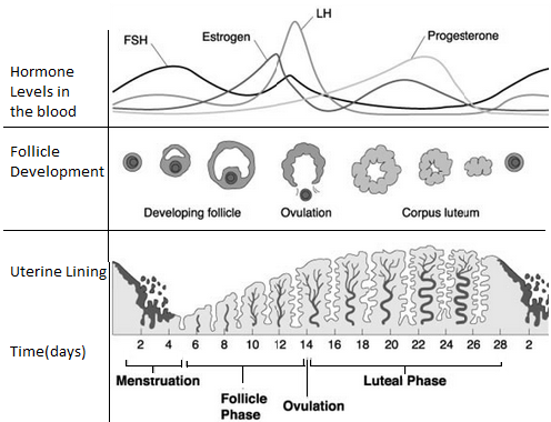
Fig: Interaction between hypothalamus, pituitary gland, and ovary showing feedback mechanisms.
Phases of Menstrual Cycle
- Follicular phase: Increased FSH and LH; follicle development.
- Ovulation: Secondary oocyte release controlled by LH.
- Luteal phase: Corpus luteum development; progesterone and estrogen secretion.
- Menstruation: Progesterone withdrawal; endometrial shedding.
Differences Between Oestrus and Menstrual Cycle
| Oestrus Cycle | Menstrual Cycle |
|---|---|
| Common in lower mammals. | Characteristic of primates. |
| Endometrium absorbed if no conception. | Endometrium shed via menstruation. |
| Sexual activity only during oestrus (heat). | Sexual activity possible anytime. |
| Heightened sexual activity coincides with ovulation. | Menstruation is prominent event. |
| Occurs less frequently (e.g., yearly). | Occurs monthly. |
Note: Humans have concealed ovulation with no obvious external signs; sexual activity peaks before ovulation.
Similarities Between Oestrus and Menstrual Cycle
- Both involve recurring physiological changes induced by reproductive hormones.
- Both start at puberty and continue until menopause.
Metamorphosis
Definition: Change in form during development.
- Controlled by brain and endocrine glands: corpus allatum, corpus cardiacum, and prothoracic gland.
- Brain: Neurosecretory cells secrete brain hormone (BH) influencing ecdysone secretion.
- Corpus Allatum: Secretes juvenile hormone (JH) controlling larval growth and molting.
- Corpus Cardiacum: Stores and releases brain hormone.
- Prothoracic Gland: Secretes ecdysone controlling pupation and adult emergence.
Types of Metamorphosis
- Complete (Holometabolous) Metamorphosis
Four stages: egg, larva, pupa, adult.
Example: Housefly, butterfly.
Larvae differ greatly from adults and specialize in feeding; pupae reorganize tissues to form adults.
Incomplete (Hemimetabolous) Metamorphosis
Three stages: egg, nymph, adult.
Example: Grasshopper, cockroach, locust.
Nymph resembles adult but molts through instars before maturity.
Advantages of Metamorphosis
- Allows juvenile and adult forms to occupy different habitats, reducing competition.
- Enables specialization of larval and adult stages for feeding and reproduction respectively.
Reproduction in Flowering Plants
The flower is the reproductive structure.
Gametogenesis in Flowering Plants
Formation of microspores (male) and megaspores (female).
Microsporogenesis produces pollen; megasporogenesis produces embryo sac.
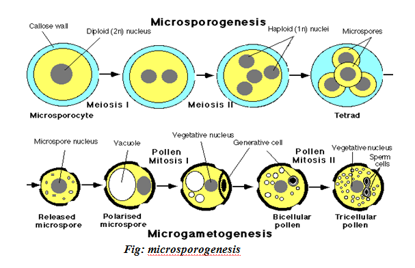
Development of Pollen Grains: Microsporogenesis
Occurs in anther pollen sacs. Pollen mother cells (2n) undergo meiosis I to form two haploid cells (dyad), then meiosis II to form four haploid cells (tetrad).
Cells separate, develop thick walls (exine and intine), and nuclei divide mitotically into generative and pollen tube nuclei.
Sexual Reproduction in Plants
Occurs in gametophyte generation within flowers.
Gametogenesis
- Microsporogenesis
- Megasporogenesis
Microsporogenesis
Microspore mother cells undergo meiosis producing microspores that develop into pollen grains (male gametophytes).
Pollen grains have two walls: inner intine and outer exine with sculptured pits.
Megasporogenesis
Occurs in ovule of ovary. Megaspore mother cell (2n) undergoes meiosis producing four haploid cells; only one develops into embryo sac.
- Embryo sac nucleus divides mitotically to produce eight nuclei arranged into antipodals, polar nuclei, synergids, and egg cell.
- Embryo sac is female gametophyte.
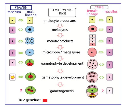

Fig: Carpel at fertilization showing ovule with diploid parent tissue and haploid embryo sac.
Double Fertilization and Its Consequences
Unique to angiosperms, involving two nuclear fusions:
First: Male gamete nucleus fuses with female gamete to form diploid zygote.
Second: Second male gamete fuses with diploid polar nuclei to form triploid primary endosperm.
Mechanism of Double Fertilization
Pollination precedes fertilization. Pollen grain lands on stigma, style secretes sugary solution absorbed by pollen, causing swelling and pollen tube growth.
- Generative nucleus divides mitotically into two male gametes.
- Embryo sac contains ovum and diploid polar nucleus.
- Pollen tube grows toward embryo sac guided by chemical signals.
- Upon reaching micropyle, pollen tube bursts, releasing contents.
- Haploid male gamete fuses with ovum forming diploid zygote.
- Second male gamete fuses with diploid polar nucleus forming triploid endosperm nucleus.


5 Comments