
SECOND TERM E-LEARNING NOTE
SUBJECT: BIOLOGY CLASS: SS 2
SCHEME OF WORK
WEEK TOPIC
- Tissue and Supporting System
- Tissue and Supporting System (cont’d)
- Mammalian Teeth
- Digestive System
- Digestive System (cont’d)
- Transport System
- Circulatory System
- Transport System in Higher Plants
- Respiratory System
- Respiratory System (cont’d)
REFERENCES
- Modern Biology for Senior Secondary Schools by S.T. Ramalingam
- Essential Biology by M.C Michael
- New School Biology by H. Stone and Cozen
- SSCE Past Questions and Answers
- New System Biology by Lam and Kwan
- College Biology by IdodoUmeh
- UTME and Cambridge Past Questions and Answers
- Biology Practical Textbook
WEEK ONE & TWO
TISSUE AND SUPPORTING SYSTEM
Contents
- Introduction
- Forms of Skeleton
- Types of Skeleton
- Functions of Skeleton
- Support in Vertebrates
- Axial and Appendicular Skeleton
- Supporting Tissues in Plants
INTRODUCTION
To carry out life processes, all organisms (plants and animals) need tissues. Tissues are group of similar cells that carry out specific functions. Skeleton is the framework of the body which provides support, shape and protection to the soft tissues and organs in animals. It forms the central core of human body and it is covered by muscles and blood vessels and skin.
FORMS OF SKELETAL MATERIALS
There are 3 forms of skeletal materials found in animals .These are
- Chitin
- Cartilage
- Bones
CHITIN
It is a tough non-living material present in arthropods (invertebrates). It acts as a hard outer covering to the animal and is made up of series of plates covering or surrounding organisms. Chitin is very tough, light and flexible. However, it can be strengthened by impregnation with ‘tanned’ proteins and particularly in the aquatic crustaceans like crabs, by calcium carbonate.
CARTILAGE
This is a tissue present in skeleton of complex vertebrates. Cartilage consists of a hard matrix penetrated by numerous connective tissue fibres. The matrix is secreted by living cells called chondroblasts. These later become enclosed in spaces (lacunae) scattered throughout the matrix. In this condition the cells are termed chondrocytes. It acts as a shock absorber in between bones during movement because it is tough and flexible with a great tensile strength. It is found predominantly in mammals and cartilaginous fishes e.g. shark.
EVALUATION
- What is skeleton?
- (a) State two main components of skeleton. (b) Differentiate between cartilage and chitin.
TYPES OF CARTILAGE
Cartilages are of three main types in mammals and they are
HYALINE CARTILAGE
This contains a dense meshwork is the most common type and can be found on surface of moveable joint, trachea and bronchi (for ease of respiration) and also in protruding parts of the nose.
WHITE FIBROUS CARTILAGE
Tougher than the hyaline cartilage and can be found in the intervertebral disc of vertebral column.
YELLOW ELASTIC CARTILAGE
Found in the external ear (pinna) and epiglottis (*cartilaginous flap covering the trachea active during food swallowing).
BONE
This is the major component of skeletal system and it consist of living cells (osteocytes), protein fibers (collagen), and minerals such as calcium carbonate and calcium phosphate. These minerals (the non- living constituent) makes up two-third of a mass of bone .Hence, bone is strong and very rigid unlike
cartilage .Bones are highly vascularised.
The skeleton of a young vertebrate embryo is made up of cartilage. As the embryo grows bone cells (osteocyte) replaces cartilage cells.Hence, the cartilage tissue becomes hardened into bone through the addition of minerals in a process called OSSIFICATION
Differences Between Bones and Cartilage
Bone | Cartilage | |
1 | Bones produce red and white blood cells | Cartilages do not |
2 | Made up of both living cells and dead cells | Made up of mainly living cells. |
3 | Bones are often rigid | Cartilage are often flexible |
4 | They are made up mainly of mineral substance such as calcium | Mineral substance are absent |
5 | Can never be replaced by cartilage | Can be replaced by bones |
6 | Flexible only in young ones | Cartilage both in young ones and adult is flexible. |
EVALUATION
- With examples differentiate between hyaline and elastic cartilage.
- Distinguish between bone and cartilage.
TYPES OF SKELETON
The three main types of skeleton in animals are
- Hydrostatic skeleton: Thisis the type present in soft bodied animals e.g. earthworm, sea anemones etc. Such animal use pressure to support itself. They also have a muscular body wall which is filled with fluid. The fluid presses against the muscular wall causing them to contract and exerting force against the fluid.
- Exoskeleton: This is the outer skeleton present in arthropods. It is secreted by the cells covering the body of the animals and the main component is chitin (non livingsubstance). Exoskeleton also supports animals against gravity and enables them to move about. Animals with these skeleton types periodically shed the old skeleton; grow rapidly in size when the new exoskeleton is still soft and extensible. The shedding process is called MOULTING or ECDYSIS.
- Endoskeleton:This is an internal skeleton present in all vertebrates. Endoskeleton of vertebrates are composed mainly of bones and the bones grow steadily as the animal grows (hence no need for moulting). Bones of many sizes and shapes make up the endoskeleton of vertebrates. These bones are attached together as moveable joints by tough flexible fibers called ligaments hence the skeleton is flexible. Muscles are also attached to the bones usually bytendons to provide posture and bring about body movement.
FUNCTION OF SKELETON
- It supports the body of organisms.
- Skeleton acts as the framework of the body
- Protection of delicate organs e.g. heart, brain, etc.
- Used for locomotion through the limbs in action.
- Important component of respiration e.g. breathing involve active movement of the ribs.
- Production of blood via bone marrows.
EVALUATION
SUPPORT IN VERTEBRATES
The skeleton of vertebrate such as fish, frog, lizard, bird and man consist of bones and cartilages. It can be classified into two.
- AXIAL SKELETON- which consists of the skull, ribs, sternum and the vertebral column.
-
 APPENDICULAR SKELETON – it is made up of limb girdles (pectoral and pelvic girdles), and the limbs (the fore and hind limbs).

AXIAL SKELETON
The Skull
The Skull is made up of flat bones joined together by suture joint which has three parts: Cranium (brain-box), facial skeleton and the jaws; including maxilla (upper) and mandible.
Functions
- It protects the brain.
- Also protects the olfactory organ, eyes, middle and inner ear.
- Gives shape to the head.
- Bears the teeth.
The vertebral column
It forms the back bone, protecting the spinal cord. It is made up of 5 groups of bones called the vertebrate each of which is built on similar basic pattern. The vertebrate are held together with strong ligament and comprehensible cartilage pads called into intervertibratal disc.
Types of Vertebrae and Location
Vertebrae | Location | Rat | Rabbit | Cat | Cow | Humans |
Cervical | Neck | 7 | 7 | 7 | 7 | 7 |
Thoracic | Chest | 13 | 12-13 | 13 | 13 | 12 |
Lumber | Upper trunk | 6 | 6-7 | 7 | 6 | 5 |
Sacral | Lower trunk | 4 | 4 | 3 | 5 | 5 |
Caudal | Tail | 30 | 16 | 18-25 | 18-20 | 4 |
A TYPICAL VERTIBRA
A typical vertebra has the following structural features
- Neural canal which is for the passage of the spinal cord.
- Neural spine which projects upward and backward for the attachment of muscle.
- Transverse processes for the attachment of muscles and ligaments.
- Centrum; a solid bony pieces below the neural canal
- Zygapophyses are the particular surfaces for joining together of successive vertebrate.
Thiscould be pre-zygapophysis (facing inward and upwards) or post-zygapophysis (facing outward and downwards
Cervical Vertebrae
The first cervical vertebra is called the atlas while the second is called the axis.
The Atlas It has a large neural canal, flat and broad transverse processes, short neural spine which could be absent at times. It also has a vertebrarterial canal for the passage of blood vessels. Centrum is absent.
EVALUATION
1. Give two classes of skeleton.
2. List types of vertebrae.
Function of Atlas
Permits the nodding of the head.
The Axis
It has a broad and flat Centrum, a large and flat neural spine, reduce transverse processesand a vertebrarterialcanal. It articulates with the atlas through odontoid process
Functions
- It permits the turning or twisting of the head.
- Forms pivot joint with the atlas.
Thoracic Vertebra
Have a long and prominent neural spine, a pair of short transverse processes, a large neural
canal and neural arc and large cylindrical centrums .They also have particular surfaces for attachment of the ribs.
Function
- Aids attachment of ribs
- Assist in breathing
- Attachment of muscles at the shoulder and back
Lumbar Vertebrae
Each has large and flat transverse processes, broad and flat neural spine, large and thick centrums and well developed zygapophyses.It has extra paired projections namely
- anapophysis
- metapophysis
Functions of Lumbar
- It provides attachment for abdominal muscles
- It bears considerable weight of the body
Sacral Vertebrae
This fuse together to form a singular bony mass called sacrum. Each sacral vertebrate has a narrow neural canal, reduced neural spine and large centrums. The first differs from the remaining four by
- Having a pair of transverse processes which is large and wing-like while the others are attached to the muscles of the back.
- Presence of a small neural canal which generally becomes narrower in the lower four vertebrae.
FUNCTION
- Joins the pelvic girdle to provide support,rigidity and strength.
Caudal vertebrae
These are joined together to form a singular bony mass called coccyx. Each has no neural spine, no neural canal and no transverse process. It appears as a solid rectangular mass of bone
Functions
- Supports the tail
- Provides attachment for tail muscle
Evaluation
- List the structural features of a typical vertebrate
- Using the location, structural features and functions, differentiate between atlas and axis.
The Appendicular Skeleton
Pectoral girdle: found around the shoulder in man and it consists of two halves which are held
by muscles. Each halve is made up of 3three bones
- Scapula
- Clavicle
- Coracoids
The scapula and coracoids are fixed together as the scapula is flat and triangular with a hollow called GLENOID CAVITY at its tip. This cavity articulates or joins with the head of humerus to form the shoulder joint.The clavicle is a small rod of bone attached to a ligament joining the sternum to the scapula
Functions
- The pectoral girdle gives attachment to muscles and ligaments.
- It provides firm support to the fore limbs.
Pelvic girdle: found around the waist in man and it consists of two halves which are joined to each other ventrally and to the sacrum dorsally. Each halve of the pelvic girdle is made up of 3threebones. They are
- Illium
- Ischium
- Pubis
These three bones form a depression (on their outer surface) called ACETABULUM which articulates with the head of the femur to form the hip joint.
Evaluation
- describe the limb girdle found in the shoulder region of the human body
- Differentiate between pectoral and pelvic girdle.
LIMBS
The limbs include the fore (upper) and the hind (lower) limbs. In most vertebrates, both limbs have the same basic plan i.e. each limb has a long bone followed by a pair of two long bones next to this is a set of small bones terminating with five digits.
The fore limbs– This is made up of an upper arm bone called humerus which joins with two other long bones at its lower end (radius and ulna) to form the elbow joints. Radius and ulna (the ulna is longer) are the bones of the fore arm, next are the wrist bones called carpals which are a small bones. These are followed by the digit bones called metacarpals which terminate in the phalanges (finger bones). In man, each digit has three phalanges except the thumb which has two phalanges.
The hind limbs-This is made up of thigh bones called femur (which is the largest and longest bone in the body). Its round upper end is the end that terminates at two rounded projections called condyles which forms the knee joint together with tibia. A small flat bone called patella is found in front of the knee joint.Next to the femur are tibia and fibula-Tibia is longer and larger. These are followed by bones of the ankle called tarsals. The lower limb terminates as at the digit bone metatarsals and each digit is made up of three phalanges
Evaluation
- on what formation plan are the upper and lower limb based
- Differentiate between the long bones of the arm and that of the thigh.
The ribs
These are long semi circular rods which connects the thoracic vertebrates to the sternum. They are found in the chest region of the body. In man, they are 12 pairs
Function
- They form a cage protecting the lungs and the heart
- They assist in breathing.
A TYPICAL RIB
A typical rib has a head, a neck and a body.The first sevenribs are connected directly to the sternum through coastal cartilages. They are therefore called true ribs. The next five are called false ribs. The eighth to tenth ribs have a common articulation to the sternum, each one attached to the coastal cartilage to the one above .The eleventh and twelfth pairs of ribs are called floatingribs because they have no connection to the sternum.
EVALUATION
- Describe the bone connecting the thoracic vertebra to the sternum.
- Classify the twelve types of ribs.
SUPPORTING TISSUES IN PLANTS
The needs for supporting tisues in plant are for:
1. definite shape;
2. strength;
3. rigidity;
4. resistance against external force such as wind and water.
Types of Supporting Tissues
Parenchyma Tissues
They are made up living cells with cellulose and many air spaces between within them. This is the most common and abundant plant tissue.
Functions
- It gives firms and turgidity to the stems of hibiscus
- stores food and water
- takes part in food synthesis in leaf mesophyll
Collenchyma Tissues
Made up of living cells which are elongated and thickened at the corners.
Functions
- Provides strength and support in young growing plant.
- Gives flexibility and resilience to plant.
Schlerenchyma Tissues
They are made up of thick cells containing cellulose and ligion.The tissues are rich in fibers.
Functions
- gives flexibility to plant
- provides strength, rigidity ,hardness and support to plant
Xylem
Xylem tissues are found in vascular tissues of stems,roots and leaves
Functions
- provides support strength and shape to the plant
- Helps to conduct water and mineral salt from the roots to leaves.
Phloem Tissues
Also located in the vascular bundles of all plants in their roots, stems and leaves
Functions
- Conduction of manufactured food from site of production to site of consumption and storage.
- Assist to provide support to the entire plant.
GENERAL EVALUATION
- Describe the structural features of a typical vertebra
- Define ossification.
- What is moulting?
- State four reasons for presence of supporting tissues in plant
- List supporting tissues found in plant and state their functions.
Reading Assignment
College Biology by IdodoUmeh. Chapter14, page 251-259
WEEKEND ASSIGNMENT
SECTION A
- …………. is a non living skeletal material
A. chondroblasts B. osteocyte C. elastic cartilage D. chitin
- The articulating surface for joining together successive vertebrates is called
A. neural spine B. zygapophyses C. transverse processes D. neural canal
- The canal for the passage of blood vessels in vertebrae is called
A. neural canal B. cervical canal C. vertebraterial canal D. zygaphosis
- Endo- skeleton is present in the following animals except
A. dog B. snake C. shark D. lizard
- The most abundant supporting tissue in plants is
A. sclerenchyma B. parenchyma C. xylem D. phloem
SECTION B
- (a) What is ecdysis? (b) Mention two animals in which it occurs
- Differentiate between (a) Bone and cartilage (b) Atlas and axis vertebrae
WEEK THREE
MAMMALIAN TEETH
CONTENT
- Types of teeth
- Structure of a tooth
- Dentition and dental formula
- Adaptation of dentition to mode of feeding
TYPES OF TEETH
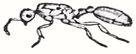
Human beings have two sets of teeth in their lifetime. The first set is called the milk teeth that grow when the child is few months old. They are about twenty (20) in numbers. These include only three types of teeth i.e. Incisors, canine and premolars. They later fall off to be replaced by the second set of teeth that consists of thirty-two permanent teeth. These are found in adult mammals. They are usually of four types: Incisors, canine, premolars and molars. These permanent teeth remain till old age.
The teeth in mammals are of four major types:
Incisors: Flat, chisel-shaped with a sharp edge. They are used for biting food, cutting and holding onto a prey to prevent its escape.
Canine: similar to the incisors. They are sharp and pointed at the tips. They are used for tearing flesh and for catching a prey.
Premolars: They are large with rigid flat surfaces which are used for chewing food.
Molars: They are used for grinding and chewing food together with the premolar.
EVALUATION
- Mention four types of teeth in human and their functions
- Differentiate between the two sets of teeth in human
STRUCTURE OF A TOOTH
The mammal’s tooth is divided into three regions
- The crown which projects above the gum
- The root which is embedded in the jaw bones.
- The neck which is the junction between the crown and the root.

The centre of the tooth consists of a pulp cavity which contains blood vessels and nerves. These are extremely sensitive to heat and cold. A layer of dentine encloses the pulp. Dentine is a hard and bone-like material which contains some living cytoplasm. The enamel is a white layer covering the dentine and is the hardest material made by animals. Enamel protects the pulp and the dentine. A layer of cement covers the dentine in the root region.
EVALUATION
- List four major parts of the mammalian tooth.
- State their functions
DENTITION
Dentition is the number, arrangement and conformation of teeth in an organism. It is concerned with the number and kind of teeth present in the mouth of an organism e.g. mammals. The teeth are arranged and fixed to the lower and upper jaw bones.
Dentition is basically of two types
Homodont dentition: that in which organisms have the same type of teeth. No set of teeth is specialized for any function. All the teeth are of the same shape, size and function. This dentition is found in fishes, amphibians and reptiles.
Heterodont dentition: In this type, organisms have teeth of different shapes, sizes and functions. This observed in mammals.
Mammals, generally speaking, have four different types of teeth, namely; incisor, canines, premolars and molar. The type of teeth possessed by an animal is closely related to the type of food it eats.
DENTAL FORMULA
A formula expressing the number and kinds of teeth possessed by a mammal. A dental formula is usually written in the form of four ‘fractions’, one for each type of tooth, with the upper and lower lines describing the upper and lower jaws respectively. Different mammals have different dental formula depending on their diet. Examples include
Man………….2[ I=2/2, C=1/1, P=2/2, M=3/3] = 32.
Dog …………… 2[ I=3/3, C=1/1, P=4/4, M=2/3] = 42.
Cow …………….2[ I=0/3, C=0/1, P=3/3, M=3/3] = 32.
Lion…………… 2[ I=3/3, C=1/1, P=3/2, M=1/1] = 30.
Rabbit …………… 2[I= 2/1, C= 0/0, P=3/2, M=3/3] =28.
Sheep …………….. 2[I=0/3, C=0/1, P= 3/3, M= 3/3] =32.
Rat …………….. 2[I=1/1, C= 0/0, P=0/0, M=3/3] = 16

GENERAL EVALUATION
- What is dentition?
- Describe dentition in a named omnivore, carnivore and herbivore.
- Mention two types of dentition and give examples of organisms that possess each.
- List four types of teeth in mammals and their functions.
- With the aid of a labeled diagram, describe the structure of a tooth.
READING ASSIGNMENT
College Biology, chapter 6, page 120 – 124
WEEKEND ASSIGNMENT
SECTION A
- The dental formula 2[I 3/3, C 1/1, PM 4/4, M 2/3] is that of
A. goat B. dog C. rabbit D. man
- Which of these teeth is usually absent in an herbivorous animal?
A. Incisor B. Canine C. Molar D. Premolar
- The type of teeth used for cutting and biting food is
A. Premolar B. Molar C. Canine D. Incisor
- Diastema can be found in the following animals except
A. Sheep B. Rabbit C. Cat D. Cow
- The hardest part of the tooth that protect the inner part is known as
A. Dentine B. Enamel C. Pulp D. Cement
SECTION B
- Describe the role of teeth in mammalian nutrition.
- State five ways of caring for the teeth.
WEEK FOUR AND FIVE
DIGESTIVE SYSTEM
CONTENT
INVERTEBRATES
- Feeding in Hydra
- Digestion in Planaria
- Digestion in Earthworm
- Digestion in insects (Grasshopper)
VERTEBRATES
- Digestion in Birds
- Digestion in Rabbits
FEEDING IN HYDRA
Hydra feeds on small aquatic organisms e.g. crustaceans. It captures its food through its structure called nematocysts found on its tentacles. The food is then passed into the mouth while the digestive enzymes are secreted into the enteron by the gland cells. The digestion is extra cellular and the digested food is absorbed into the body while the undigested food is sent back into the enteron and released through the mouth. The digestion here is therefore intracellular and extracellular.

DIGESTION IN FLATWORM [TAPEWORM & PLANARIA]
Tapeworm is a parasitic worm which has no alimentary canal; it feeds on digested food of the host. However, the planarian is a free living flatworm which feeds on small animals. Planaria has a simple alimentary canal with one opening i.e. the mouth present in the ventral part of its body cavity. The pharynx of the planarian can be extended out of the mouth during feeding. The pumping action of the pharynx sucks in pieces of food which enter the pharynx and move into the small intestine. Digestion is partly extracellular and partly intracellular. The branched intestine enables the digested food to diffuse to all parts of the body while undigested food is egested through the mouth.

EVALUATION
- Compare feeding in hydra with feeding in planaria
- Explain extracellular digestion.
DIGESTION IN EARTHWORM
The alimentary canal of the earthworm is a tube with two openings:
- The mouth through which the food enters
- The anus through which the undigested food leaves the body
The alimentary canal of the earthworm has the following parts:
- Mouth and thin walled buccal cavity for food ingestion
- A muscular thin walled pharynx which secretes mucus to lubricate food particles
- A narrow tubular thin walled oesophagus
- A thin walled crop for storing food
- A thin walled muscular gizzard where food is ground against small stones to break it up.
- A long straight intestine in which extracellular digestion occurs.
MECHANISM OF DIGESTION IN EARTHWORM
The prostomium is the mouth of the earthworm. Since it has no jaws or teeth, the earthworm uses its muscular pharynx to suck in soil containing food. The food particles and soil go through a long esophagus into a round organ called a crop. The crop stores the food temporarily. Then it is forced into a very muscular organ called the gizzard. The gizzard contracts and expands, causing grains of sand and food to rub together. In this way, the food is ground up. Food is digested in the intestine, which stretches from segment nineteen to the end of the worm. Here enzymes chemically break down the food. Then the digested food is absorbed by the blood circulating through the intestinal walls. The earthworm’s digestive system can be thought of as a tube within a tube.
The complicated organs of the digestive system take up most of the anterior half of the earthworm’s body. The earthworm ingests large amounts of soil, which has organic matter in it. The useless inorganic matter goes through the worm with no change. This is often left on the surface of the ground in the form of castings. As the earthworms feed, they loosen the soil, making it easier for air and water to enter. Their wastes also add to soil fertility. If there were no earthworms, the soil could not support crops, grasses, and other plants.
EVALUATION
- Mention all the parts of earthworm involved in digestion.
- Explain how castings are formed in earthworm.
DIGESTION IN INSECTS
The alimentary canals of insects consist of three major parts:
- The fore gut [mouth, pharynx, oesophagus crop, gizzard]
- Mid gut [stomach]
- Hind gut [intestine, ileum, colon, rectum, anus]

Insects such as grasshoppers feed on leaves. They use their mouth parts to cut and crush the leaves. Saliva is introduced or poured into the leaves from the salivary gland. The saliva helps to soften the leaves and the chewed food is in the crop, and broken up further into small pieces.
The foregut and the midgut secrete enzymes rich juice into the midgut where digestion and absorption occurs.
The hindgut is for water absorption. Only solid faeces pellets are egested from the anus after the food waste has been collected through the malpighian tubules joining the mid and hindgut.
DIGESTION IN BIRD
Birds do not have teeth but horny beak which they use for feeding. In many birds, the feet also show adaptation for feeding.
The alimentary canal of the bird consists of the following:
1. Oesophagus 2. Crop 3.Proventriculus
4. Gizzard 5. Small intestine 6. Caeca 7. Cloaca

MECHANISM OF DIGESTION IN BIRDS
The bird swallows their food whole and store in the crop. In the crop, it is softened by secretion from the wall of the crop. It is then through the proventriculus to the gizzard where gastric juice churns the food and breaks it up into smaller units. This is preceded by the grinding action of the muscles of the gizzard [a strong muscular bag]. Small stone in the gizzard also assists in the grinding of the food.The digestion is completed in the small intestine by the action of intestinal and pancreatic juice. The absorption also occurs here and the solid waste passed through the anus into the cloaca.
EVALUATION
- State the three parts that make up the digestive system of insects.
- Describe the mechanism of digestion in bird.
DIGESTIVE SYSTEM AND DIGESTION IN MAN
Heterotrophic organisms get their food from their autotrophic counterparts or depend on other heterotrophic organisms.
The food is taken in (ingestion), broken down into simple, soluble and diffusible substances through some chemical and mechanical processes. This is referred to as digestion. The digested food is eventually absorbed (absorption) into the body fluids and assimilated while the undigested food is removed (egested)
FEEDING AND DIGESTION MECHANISMS
All holozoic animals have structures for obtaining their food. The structures also help them to capture their preys. Those organisms feeding on large food particles have their bodies modified into structures like claws, teeth, beak etc. However, those feeding on small pieces of food have either fluid feeding or filter feeding structures while saprophytes change their food to digested absorbable form before taking them in. Parasites on the other hand have structures for boring into the bodies of their host.
Most holozoic animals have a digestive pathway called alimentary canal or gut unlike unicellular animals. Their gut consist of two openings
- Anterior opening called mouth
- Posterior Opening called anus
A typical alimentary canal is adapted for breaking food into smaller units, producing digestive secretions and absorbing digested food and water.
A digestive system is made up of alimentary canal and the associated glands and organs which produce some of the enzymes-rich secretion that bring about digestion.The action of the teeth is the mechanical breakdown of digestion while the digestive enzymes speed up the chemical digestion.
DIGESTION OF FOOD IN HUMAN
Food passes through the following process in man
Ingestion →Digestion →Absorption →Assimilation →Egestion
Food is ingested in the mouth and the teeth grind the food into smaller units, chemical digestion also begins. Saliva contains an enzyme, ptyalin that acts on cooked starch to convert it to complex sugar (maltose). Saliva is a watery, slightly alkaline substance secreted by the salivary gland.
The tongue mixes the food with saliva and rolls it into a ball (bolus) which is then swallowed. The food passes down into the stomach through the gullet (oesophagus). During swallowing of food, the entrance to the trachea must be closed to prevent choking. The wall of the esophagus is muscular and it contracts and relaxes to push each bolus of food downward, this process is called peristalsis.
DIGESTION IN THE STOMACH
The muscular wall of the stomach contracts and relaxes forcefully just churning the food. The gastric juice mixes with the food. Gastric juice contains two important enzymes; pepsin and rennin as well as hydrochloric acid for activating pepsinogen into pepsin. Pepsin digests protein into peptones and polypeptides which are intermediate products in protein digestion. Pepsin works best in acidic medium and the acid also assists to kill the bacteria present in food.Rennin causes the coagulation of milk into thick curd (convert soluble caseinogens to insoluble casein). Food stays in the stomach for about 3-4 hours.
DIGESTION IN THE SMALL INTESTINE
The first part of the small intestine is duodenum; the pancreas secretes pancreatic juice which contains digestive enzymes. The liver produces bile, which is stored in the gall bladder. Bile is a greenish liquid that emulsifies fat, which does not contain digestive enzymes.
The pancreatic juice contains three important enzymes:
- amylopsin converts starch to maltose
- Trypsin converts protein to polypeptides
- Lipase converts Lipids to fatty acids and glycerol

The latter part of the small intestine is the ileum. Here the wall of the intestine secretes five important enzymes:
- maltase – converts maltose to glucose + glucose
- Sucrase – converts sucrose to glucose + fructose
- Lactase – converts lactose to glucose + galactose
- Erepsin – converts polypeptides to amino acids
- Lipase – converts fats and oil to fatty acids and glycerol
In man the digestion of food ends in the small intestine. Hence the end product of protein is amino acids, fats and oil is fatty acid and glycerol while that of starch are glucose, fructose and galactose.
ABSORPTION AND ASSIMILATION
Glucose, amino acids, fatty or carboxylic acids and glycerol as well as vitamins and mineral salt are absorbed in the small intestine. For efficient absorption, a large surface area is needed. To ensure this, the wall of the small intestine has folds and furrows. Also there are finger-like projections called villi (Villus). The inner surface layer (epithelium) of each villus is thin. This allows the absorption of the end products of digestion which takes place by either diffusion or active transport. The absorbed food substances are carried away through the blood vessels and lymphatic vessels (containing blood and lymph respectively).
In each villus, there is a blind lymphatic tube called lacteal which is surrounded by a network of blood capillaries. The lymph in the lacteal transport fatty acid (carboxylic acid) and glycerol which recombines to form fats in the lacteals. This is then carried by the blood to where they are needed. Excess fats are stored in fat cells to form adipose tissues which are usually found under the skin and around organs.
EVALUATION QUESTION
- Why are humans heterotrophic?
- Explain the mechanism of digestion after taking a plate of rice and beans.
- FILTER FEEDING: This concerns mainly aquatic animals which feed on very tiny organisms in water. They use their sieve like structure to collect their food or prey.Examples of filter feeders are mosquito larva, ducks, prawns etc.
- FLUID FEEDING:This concerns animals which feed on fluid materials and so they are called fluid feeders. They include classes of animals namely:
(a) Sucker e.g. bugs, mosquitoes, butterfly, housefly, tsetse fly etc.
(b) Wallowers:These are organisms which wallow in their food e.g. tapeworm. Tapeworm lives within the digested food of its host and absorbs the food directly into the body. Therefore, it does not have alimentary canal. The absorption of its food is through its entire body surface.
MODIFICATION AND MECHANISMS OF FEEDING IN ANIMALS
This can be studied in five mechanisms:
- absorption mechanisms e.g. tapeworm
- Biting and chewing mechanisms e.g. grasshopper
- Sucking mechanisms e.g. mosquitoes
- Grinding mechanisms e.g. Man
- Trapping and absorbing mechanisms e.g. bladder worm.
GENERAL EVALUATION
- State the various feeding habits animals.
- Give two organisms each for the above listed habits
- What is an enzyme?
- Draw the longitudinal section of a villus.
- Summarize digestion in a tabular form under the following columnar topics (a) site of digestion (b) juice secreted (c) site of secretion of juice (d) enzymes produced (e) digestion process carried out by enzymes.
READING ASSIGNMENT
College Biology, Chapter 6, Page 106 – 131
WEEKEND ASSIGNMENT
SECTION A
- Digestion (breaking down of food into simple soluble and diffusible forms) occurs by ……… and ……… processes
- The process by which digested food is absorbed into the body cells is called
A. ingestion B. egestion C. assimilation D. digestion
- Chemical digestion of starch starts in the …………
A. intestine B. Mouth C. stomach D. pancreas
- The enzyme involved in the coagulation of milk into thick curds is ………
A. ptyalin B. pepsin C. Rennin D. Lipase
- The end product of starch digestion is ……..
A. glucose B. maltose C. glycogen D. amino acids
SECTION B
- State the region in the alimentary canal where the chemical digestion of the following food
substances begins(a) Carbohydrate (b) Protein (c) Lipids
- What role does pancreatic juice play in digestion?
WEEK SIX
TRANSPORT SYSTEM
CONTENT
- Transport system
- Need for transport system
- Relating transport in lower organism to that in higher organism
- Transport material in animals and plant
- Diverse mechanism of transportation in some organisms
- Transport system in mammals (man)
- Functions of blood
Transport system
Transport system is the movement of metabolic materials from various parts of an organism where they are produced and transported to other parts where such are used, stored or removed from the body.
Need For Transport System
All living organisms (plants and animals) need transport system for the following reasons
- To obtain essential materials such as oxygen, water and nutrients.
- To remove metabolic waste such as carbon dioxide, urea, water, etc.
- For moving water and mineral salts from the soil through the roots to the various parts of plant.
- For transfer of hormones from production site to site of action.
- For transfer of glucose to various parts of plants.
RELATING TRANSPORT IN LOWER ORGANISMS TO THAT IN HIGHER ORGANISM
Transport in Lower Organisms | Transport in Higher Organisms | |
1 | Substances are moved over small distance | Substances are moved over greater distance |
2 | Transport is by simple diffusion | Transport involves diffusion and other means. |
3 | Diffusion is enough because the surface area to volume ratio (A/V) is great | Effusion transport system is necessary because surface area to volume ratio (A/V) is too small |
4 | Cells are not isolated | Isolated group of cells need to be connected |
5 | Transport materials are small in quantity. | Transport materials are large in quantity. |
EVALUATION
- What is transport system?
- State the needs for transport system.
Transport Materials In Animals
Materials Transported | Source | Destination | |
1 | Oxygen | Lungs | All living cells of the body |
2 | Carbon dioxide | Body cells | Lungs |
3 | Urea | Body cells | Liver |
4 | Excess salts | Body cells | Skin and kidney |
5 | Water | Body cell | Skin, lungs, liver, kidney etc |
6 | Amino acid | Small intestine | Body cells |
7 | Vitamins | Small intestine | Body cells |
8 | Sugar | Body cells | Body cells |
9 | Fatty acid and glycerol | Small intestine | Body cells |
10 | Mineral salt | Small intestine | Body cells |
11 | Hormones | Endocrine glands | Target organs of tissue |
12 | Antibodies | White blood cells | All body parts |
Transport Materials in Plants
Materials Transported | Source | Destination |
Manufactured food | Leaves | All body cells |
Excretory Products (C02 and water) | All living cells | Site of excretion e.g. stomata |
Water (absorbed) | Soil | Leaves and other parts of the leaves |
Other materials transported in plants are:
1. Oxygen 2.nitrogen waste products (latex) 3. amino acids 4. glucose 5. lipids 6. auxins (hormones) 7.mineral salts
Transport Media
Liquid or fluid is usually the medium of transportation of minerals. Generally speaking, the four major media of transportation in organisms are:
Cytoplasm: Used in lower unicellular organisms such as amoeba, chlamydomonas, euglena, etc.
Cell sap/ Latex: A concentrated solution in the cell vacuole of plants.
Blood: Used in most animals, especially vertebrates for conveyance of essential materials like oxygen, digested food, etc.
Lymph: Found in higher animals. Lymph is a fluid with extra lymphocytes (W.B.C with no red blood cells present). It returns its fluid to the main vein through opening in the subclavian (left jugular) vein below the neck.Lacteal is a lymphatic vessel transporting fatty acids and glycerol. The lymph movement is enhanced by muscular action. It moves through lymph vessel. Some swellings exist in the gut along the lymphatic vessel, especially in the neck, groin and armpit called lymph nodes. These are where lymph passes through to be purified before entering into the blood stream. Unlike the circulatory system, the lymphatic system ends blindly.
Evaluation
- Mention two differences between the lymphatic system and the circulatory system.
- List transport materials common to both plants and animals
Diverse Mechanisms of Transportation in Some Organisms
Unicellular Organisms
Materials are transported through the continuous streaming movements. The streaming could be along the direction of movement of the organism, back to front (e.g. Amoeba) or in circular motion (e.g. Paramecium)
Multicellular Organism
- Hydra
The movement of the gut wall draws water into the gut and causes digested food and oxygen within it to circulate. Thus the cell lining the gut absorbs the materials. The whopping movement of the flagella of flagellated cells also helps in material circulation in the gut.
- Flatworms
The large body surface area to volume ratio and extensive branching gut throughout the body makes the food and oxygen to diffuse into all the body cells. Movement of the body wall assists to transport waste products out of the body.
- Insects and mollusks
Both have open circulatory system i.e. the heart pumps blood out into a blood vessel with branches open into spaces in the body cavity known as Haemocoels. Blood from these spaces eventually flows into the vessels leading into the heart. Blood flows in unidirectional and blood distribution is poorly controlled.
TRANSPORT SYSTEM IN MAMMALS (MAN)
The media of transportation in man include the blood and lymph.
COMPOSTION AND STRUCTURE OF BLOOD
The blood is a tissue in a fluid form. It is about 5-6 liters in the body. Blood is made of two major components.The blood cells (corpuscles); which are solid.The plasma which; is liquid.

BLOOD CELLS
There are three types
- Red blood cells (erythrocyte)
- White blood cells (leucocytes)
- Blood platelets (thrombocytes)
Blood Cells | Description | Function |
RBC |
| Helps to transport oxygen from lungs to the body cells through its pigment (haemoglobin). Haemoglobin combines readily with oxygen to form oxyhaemoglobin in the lungs |
WBC |
| Help to defend the body against diseases by engulfing and intruding pathogens (bacteria and virus) or by secreting antibodies. |
RBC- Red Blood Cell WBC- White Blood Cell
White blood cells areof two types;
Phagocytes- found in lymphatic system which ingest bacteria, viruses and dead cells to prevent diseases in a process called phagocytosis.
Lymphocytes– made in lymph glands and they produce antibodies i.e. chemicals which stick to the surface of germs to kill them.
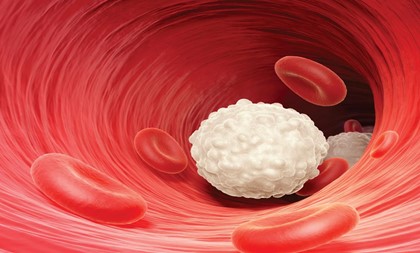

Blood cell | Description | Function |
Platelets |
| Aids in blood clotting |
Blood plasma (transport liquid) |
| Transport the dissolved substances and the blood cells. |
Lymph |
|
|
FUNCTIONS OF BLOOD
- Oxygen is transported through hemoglobin.
- Temperature regulation by evenly distributing heat produced in the liver and the muscles throughout the body.
- Transportation of digested food (glucose, amino acid, fatty acids and glycerol) from the villi to all body cells and tissues for use or storage.
- Transportation of excretory products (C02, water, urea) from site of production to excretory organs like skin, lungs, liver and kidney for removal
- Transfer of hormones from production site to target organs.
- Transportation of water (90% of the blood content) to various cells for metabolic activities.
- Defense against infection through the action of WBC
- Blood clotting initiated by the platelets when injury is sustained
- Production of anti bodies by the WBC for destroying pathogens and their harmful product.
GENERAL EVALUATION
- State the media for transportation in ten organisms.
- Compare the transport mechanism of a named unicellular organism to that of a named multi-cellular organism
- Draw the diagram of RBC, WBC and platelets (label them accordingly)
- What feature distinguishes lymph from blood plasma?
- State five functions of blood.
Reading Assignment
College Biology, Chapter 7, Page 147 – 156
WEEKEND ASSIGNMENT
SECTION A
- The following are true except one about transport in amoeba
A. substances are removed over greater distance B. transport is by simple diffusion C. diffusion is enough for transport D. substances are moved over short distance
- The following metabolic materials are transported from body cells to the skin and kidney
A.sugar B. hormones C. excess salt D. protein
- The transport medium which plays active roles in the defense and metabolism of lipids is … A. cytoplasm B. lymph C. blood D. platelets
- The following animals except one has open circulatory system
A. goat B. house fly C. snail D. grasshopper.
- One of the following has nucleus
A. red blood cell B. white blood cells C. platelets D. hydra
SECTION B
- (a) Why is blood a tissue?
(b) Differentiate between blood and lymph.
- Describe the transport system in a named mollusk.
WEEK SEVEN
CIRCULATORY SYSTEM
CONTENT
- Protective Function of Blood
- Circulatory System in Mammals
- Blood Vessels and the Heart
PROTECTIVE FUNCTION OF BLOOD
The blood performs two basic functions in mammals and
These are:
- Transportation of various substances
- Protection of body against diseases
The blood defends the body in major ways
- Antibody production (Clumping): The lymphocytes produce antibodies which are any of a large variety of proteins normally present in the body or produced in response to an antigen which it neutralizes, thus producing an immune response.This can also be ensured by injecting small dose of the weaker or dead pathogens into the body. This process is known as vaccination or immunization
- Neutralization: The whole blood will also produce antitoxins which neutralize the toxins produced by the pathogens.
- Phagocyte Action (Engulfing): Phagocytes engulf the pathogens and digest them.
- Clotting: The blood platelets clot the blood to prevent germs from entering the body and also prevent loss of blood
MECHANISM OF CLOTTING
When a blood vessel is damaged and exposed to air, platelets in the blood stream release an enzyme thrombokinase. The thrombokinase then converts prothrombin (inactive blood protein) to an enzyme called thrombin. The thrombin then converts thesoluble fibrinogen in the plasma to insoluble fibrin in the presence of calcium ions. The threadlike fibrin then forms a network or mesh on the surface of the wound and blood cells are trapped within the network or mesh to form a clot. The clot dries to scab over the wound.

EVALUATION
- Describe the mechanism of blood clotting.
- What is vaccination?
TYPES OF CIRCULATORY SYSTEM
The circulatory system in animals can be:
- Closed or open
- Single or double
Closed Circulatory System: This involves the blood vessel called arteries which divides capillaries which later join up with other vessels called veins. Blood is therefore limited to the vessels and the heart it does not have direct contact with the body cells this system is observed in annelids and vertebrates.
Opened Circulatory System: The blood vessels lead out of the heart but end in blood spaces called haemocoels within the body cavity. The blood has direct contact with the cells after which it is returned to the heart. Arthropods and some mollusks have open circulatory system
Single Circulatory System: The blood passes through the heart once in a complete movement round the body e.g. fishes because of their two chambered heart have single circulatory system.
Double Circulatory System: The blood passes twice in the heart every time it makes one complete movement round the body. Each time it passes through a separate path way e.g. mammals. Double circulation gives rise to pulmonary and systematic circulation.
BLOOD VESSELS
- Arteries: These are vessels that carry oxygenated blood away from the heart to the body’s organs except for pulmonary artery that carries deoxygenated blood.
- Arteriole: A branch of an artery that gives rise to capillaries.
- Veins: These are large vessels that carry deoxygenated blood toward the heart except for pulmonary vein which carries oxygenated blood.
- Venule: Small vessel that carries blood from the capillaries to the veins.
- Capillaries: They link the arteries with the veins around the tissues and organs. They are tiny and thin walled to facilitate easy exchange of gasses nutrients and waste products between the cells and the blood

EVALUATION
- Define (a) artery (b) venule (c) capillary
- What is double circulatory system?
DIFFERENCES BETWEEN ARTERY AND VEIN
ARTERY | VEIN | |
1 | It has a thick (muscular walls) | It has thin wall |
2 | Elastic wall | Non-elastic wall |
3 | Carries blood away from the heart | Returns blood to the heart |
4 | Carries oxygenated blood except pulmonary artery | Carries deoxygenated blood except pulmonary vein |
5 | Blood is pink or bright colour | Blood is dark red in colour |
6 | Situated deep in muscles | Superficially situated |
7 | Has small lumen | Large lumen |
8 | Pressure is high | Pressure is low |
9 | Pulse is readily detectable | Pulse is not easily detected |
10 | It has no valve except semi lunar valves | It has valves |
THE HEART
The heart is a muscular and powerful organ responsible for pumping blood in the system of mammals. It is located within the chest cavity and protected in the ribs and sternum. The pumping action of the heart is known as heart beat (heart beat). Heart beat per second varies from animal to animal and is often increased at moments of excitement e.g. Humans heart is about 72 beats per minutes.
The special muscles making up the heart is known as cardiac muscles and the heart is enclosed in a two layered tough protective membranes called the pericardium. Pericardial fluid fills the space between the heart and pericardium. It reduces the friction arising from pumping movement of the heart against its surrounding tissues.
STRUCTURE OF THE HEART
The human heart is divided into four chambers, the right and left auricles, the right and left ventricles. The walls of the ventricles are often thicker than those of the auricles. The left ventricles especially have a thick wall because it pumps blood out to all other parts of the body and this requires more pressure.
The heart is divided into two halves by a central barrier called septum. Bicuspid valves separate the left auricle and the left ventricle. This ensures that blood flows only in one direction i.e. from the auricles to the ventricles. Similarly the tricuspid valve exists between the right auricle and the right ventricle. It serves the same function as the former. These bicuspid valves are held in place by special fibers, non-elastic cords known as chordae tendineae.
HEAT BEAT
The heart beat consist of alternate contractions and relaxation of the right and left auricles as well as the right and left ventricle. Human heart beat is about 72 beat per minutes this can be divided into two phases, namely
- Diastole: This is the first stage of the heart beat; the two auricles contract forcing blood into the ventricles and oxygenated blood into the left ventricle. As the ventricles gets filled up, the cuspid valves are pushed up and closed.
- Systole: This is the second phase of the heart beat. The ventricles contract sending blood out to the two tracks of the main arteries and out of the heart. Deoxygenated blood from the right ventricles passes into the pulmonary artery while oxygenated blood is sent into the aorta. The sequence repeats itself.


BLOOD VESSELS AND ORGANS SUPPLIED.
Blood Vessel (Artery) Organ Supplied
- Carotid artery ……. Head
- Pulmonary artery …….. Lungs
- Hepatic artery …….. Liver
- Gastric artery …….. Stomach
- Mesenteric artery …….. Intestine
- Renal artery ……… kidney
- Gonadal artery ……… Gonads
- Intercostals artery ……… Wall of thorax
Corresponding veins accompany the arteries stated above
GENERAL EVALUATION
- Draw a well labeled diagram of the heart
- Differentiate between arteries and veins
- Explain systole and diastole.
- Draw the cross section of the artery
- What is pulmonary circulation?
Reading Assignment
College Biology, Chapter 7, Page 136 – 157
WEEKEND ASSIGNMENT
SECTION A
- The following are the basic functions of blood exceptA.Transportation of substances B. Reproduction
C. Protection of the body D. Transportation of hormones
- ………. is actively involved in preventing excessive loss of blood at injury A. Red blood cell B. White
blood cell C. Platelet D. Serum
- The following except one have closed circulatory system. A. Snail B. Earthworm C. man D. lion
- Semi lunar valve is present in A. Arteries B. Veins C. Capillaries D. Bladder
- Human heart beat is …….. per minute A. 60 B. 66 C. 72 D. 80
SECTION B
- With a well labeled diagram only, describe the structure of the heart.
- State the differences between arteries and veins.
WEEK EIGHT
TRANSPORT SYSTEM IN HIGHER PLANTS
In simple unicellular plant materials are exchanged by simple diffusion process between the plants and its aquatic environment. Hence there is no need for elaborate transport system. However, in higher plant such as ferns and the flowering plants. There is a need for elaborate transport system for transporting water and mineral salts from the soil to the various parts of the plants and also transport manufacture food from the leaves to other parts where it is either used up or stored up. The transport system of a plant is made up of vascular bundles consisting of the xylem and the phloem tissues.
The xylem is responsible for conducting water with dissolved substances from the soil to other parts of the plant. The phloem tissue is responsible for the transportation of manufactured food from the leaves to other parts of the plant (translocation). In the roots and stems of dicotyledonous plants, a layer called cambium exists between the xylem and the phloem tissues. The vascular bundles therefore are found in the roots, stems and leaves of flowering plants.
PROCESSES WHICH AID TRANSPORTATION IN PLANTS
The four processes supporting transportation in plants are:
- Translocation: The process by which manufactured food substances are transported through phloem tissue from site of production to plant tissues where they are used or stored. It is usually from leaves to other plant parts.

- Transpiration: The removal of excess water from plants into the atmosphere in form of water vapour. The loss of water can be through the stomata (in leaves) lenticels (in stem) or cuticle of the leaf surface. The rate of transpiration is measured using the instruments, potometer.
CONDITION AFFECTING THE RATE OF TRANSPIRATION
- The Size of the Stomatal Pores: Flaccidity of the guard cells causes them to close preventing transpiration but when turgidity occurs, the cells open for transpiration to take place.
- Humidity: The higher the humidity, the slower the rate of transpiration.
- Temperature: Increase in temperature leads to increase in transpiration.
- Light: High light intensity causes high photosynthetic rate which in turn leads to increase in temperature thereby causing high rate of transpiration.
- Wind: The higher the speed of wind, the higher the rate of transpiration.
- Soil Water: Higher level of soil water leads to higher rate of absorption which results in higher rate of transpiration.
IMPORTANCE OF TRANSPIRATION TO PLANT
- It helps plants to absorb water and mineral salts from the soil.
- It facilitates the movement of soil water.
- Cooling the plant after water evaporation has taken place.
- Absorption of water and mineral salt by roots of plant water from the soil enters the plant through the root hairs by osmosis. This leads to increased turgor pressure of the vacuole of root cells. The water absorbed then gets into the xylem vessel
- Water transport in the xylem tissue: This is due to the following
- root pressure and suction pressure,
- Capillary action due to alteration between the water molecules and the walls of the xylem vessel,
- transpiration pulls.
GENERAL EVALUATION
- List the three main blood vessels in human body
- Draw and write short note on the human heart
- Write short note on transportation in plants
- List four processes that aid transportation in plants.
Reading Assignment
College Biology, Chapter 7, Page 159 – 169
WEEKEND ASSIGNMENT
SECTION A
- The rate of transpiration is measured using the instrument called A. photometer B potometer C. secchi disc D. hygrometer.
- The process by which manufactured food substances are transported through phloem tissues from site of production to plant tissues where they are used is A. transpiration B. translocation C. active transport D. passive transport
- The following except one have closed circulatory system A. Snail B. earthworm C. man D. lion
- Semi lunar valve is present in A. arteries B. veins C. capillaries D. Bladder
- Human heart beat is ….. beats per minute A. 60 B. 66 C. 72 D. 80
SECTION B
- With a well labeled diagram, describe the human heart.
- Explain (a) root pressure (b) suction pressure
WEEK NINE
RESPIRATORY SYSTEM
CONTENT
- Definition / Phases of Respiration
- Conditions Necessary for Respiration
- Characteristics of Respiratory Surface
- Types of Respiratory System
- Mechanism of Respiratory System in Lower Animals & Higher Animals
DEFINITION/PHASES OF RESPIRATION
The process of respiration involves the taking in of oxygen its transport within the body of the organism, its exchange in the cells and the eventual release of energy in form of ATP, water and carbon(IV)oxide. The energy is utilized by the cells for their daily activities. Respiration can therefore be defined as a biochemical activity of the cell in which glucose is broken down in a series of reaction controlled by enzymes to release energy.
The following are the Different Phases of Respiration
- External Respiration (Breathing)
This is the taking in of oxygen (inhalation)into the respiratory organ(e.g. lungs or gills) and breathing out (exhalation) of carbon(IV)oxide and water vapour.
- Internal (Tissue) Respiration
This is the oxidation of food substances within the cells leading to the release of energy,carbon(IV)oxide, and water. This is made possible by the oxygen taken in through the breathing process. This can be represented by the following equation
C6H12O6 +6O6 →6H2O+6C02+Energy (ATP)
CONDITIONS NECESSARY FOR RESPIRATION
For effective exchange of gases to take place, the following conditions must be met:
Respiratory Medium: This refers to the environment from which the organism picks up oxygen e.g. air and water.
Respiratory Organ: This refers to the organ possessed by the organism needed to pick oxygen from the environment and pass out CO2 and water vapour e.g. lungs in mammals and gills in fishes, etc.
Transport Medium: This is needed to transport dissolved oxygen to the various cells of the body which in turn picks up CO2 and other waste productsfor elimination process e.g. blood in mammals.
Ventilation: This is the movement of air in the direction over the respiratory surface. This ensures the replacement of used oxygen and the elimination of waste products. Increase in the rate of gaseous exchange isfacilitated by ventilation mechanism e.g. breathing in human.
Respiratory surface: This refers to the actual surface of the body where gaseous exchange takes place e.g. alveoliin mammals and cell membrane in amoeba
CHARACTERISTICS OF RESPIRATORY SURFACE
The following characteristics must be exhibited by all respiratory surface be it in plant or animals
- Respiratory surface must be moistened because gases diffuse in solution through them.
- It must be permeable to allow gas to pass in and out of them.
- It must be thin-walled to make diffusion easier and faster.
- It must have adequate supply of transport medium e.g. blood.
- The surface must be large to aid easy diffusion of gases.
- It must be highly vascularized i.e. it must have lots of capillaries or similar network to bring in and take away gases.
EVALUATION
- What is respiration?
- Mention five characteristics of a respiratory surface.
TYPES OF RESPIRATORY SYSTEM
Thesevary from organism to organism depending on their type, complexity, size and habitat in which they can be found. The table below reflects various organisms and respiratory structures related to them
Organism | Respiratory Structure | |
1 | Unicellular organism e.g. Amoeba and paramecium | Body surface |
2 | Hydra worm tapeworm | Cell membrane |
3 | Earth worm | Wet skin or body surface |
4 | Fishes e.g. Tilapia | Gills |
5 | Arthropod e.g. insects | Tracheal system |
6 | Arachnids e.g. spiders | Lungs books |
7 | Tadpoles | Gills |
8 | Reptiles e.g. lizards | Lungs |
9 | Amphibians e.g. toads | Mouth, skin, and lungs |
10 | Mammals | Lungs |
11 | Flowering plant | Stomata and lenticels |
MECHANISM OF RESPIRATORY SYSTEM IN LOWER ANIMALS
Unicellular Organism
They require no elaborate respiratory system for they respire through their entire body surface (the cell membrane acts as the respiratory surface) through simple diffusion process.Oxygen which is highly concentrated in the environment is absorbed in the organism which diffuse into the region with lower concentration.
Insects
Tracheal system is what insects use for respiration. Air passes through a spiracle then enters tracheal trunk which diffuses throughout a complex, branching network of tracheal tubes that subdivides into smaller and smaller diameters and reaches every part of the body. At the end of each tracheal branch, a special cell (the tracheole) provides a thin, moist interface for the exchange of gasses between atmospheric air and a living cell. Oxygen in the tracheal tube first dissolves in the liquid of the tracheole and then diffuses into the cytoplasm of an adjacent cell. At the same time, carbon dioxide, produced as a waste product of cellular respiration, diffuses out of the cell and, eventually, out of the body through the tracheal system.
EVALUATION
- State the organs of respiration in (a) Amoeba (b) cockroach (c) spider (d) scorpion (e) tadpole
- Differentiate between the mechanism of respiration in Amoeba and cockroach
FISH
The gill is the respiratory organ in the fish. Enclosed in the gill chamber and about three to four in numbers. Each gill is made up of a gill filament (where gaseous exchange takes place) and the gill raker (that prevents food particles from entering the gill chamber) and the gill arch (in which the filaments are built). The gills are concealed by the operculum.The fish initiates breathing by closing the operculum and opening the mouth and lowering the floor of the mouth. Water which contains dissolved oxygen rushes into the mouth of the fish. The fish then closes its mouth and water rushes into the gill chamber and moves across the gill filaments. Oxygen in the water then diffuses into the gill filaments while CO2 diffuse out of the body expelled through water as the fish opens its operculum.

TOAD
Tadpole: This is a larval toad or frog that hatch from eggs and can only survive in water. Tadpoles have tiny external gill flaps that extract oxygen from water as it passes over them. Tadpoles open their mouths as they swim and take in water. As the mouth closes, muscles transfer the water to the gills. These consist of thin membranes called lamellae, which take oxygen from the water where it enters the blood stream through the process of diffusion. Tadpoles can also rise to the surface and gulp oxygen from the air. As tadpoles mature the gills are absorbed by the body as other respiratory systems develop.
Adult Toad:
The adult toad respire in three difference ways namely: the skin, mouth, and lungs
BUCCAL (MOUTH) GASEOUS EXCHANGE
The toad is able to utilize its mouth as a respiratory organ because of the following:
- It is very large i.e. has a large surface area.
- It has a thin mucus membrane for easy diffusion
- It is well supplied with blood capillaries
To initiate breathing, the toad closes its mouth, the nostrils are opened and the floor of the buccal cavity is lowered, air is drawn through the nostrils into the buccal cavity. After this, the capillaries and the glottis close and gaseous exchange takes place between the blood and the inhaled air. To get rid of air, the floor of the buccal cavity is raised increasing the air pressure, and hereby forcing the nostrils to open and air in the buccal cavity containing carbon dioxide flows out.
EVALUATION
- State the structure involved in respiration in fishes.
- Differentiate between the mechanism of respiration in fish and that of toad.
SKIN /CUTANEOUS RESPIRATION IN TOAD
This is possible because of the large surface area of the skin. The skin is moist because of continuous secretion form the mucus gland, there is adequate supply of blood capillaries and finally, the membrane serving as the skin is thin. simple diffusion of gases takes place through it both on land and water.
LUNGS /PULMONARY RESPIRATION IN TOAD
This is similar to what is obtainable in the buccal respiration but with a slight difference. In order to draw air into its mouth the toad lowers the floor of its mouth, which causes the throat to expand. Then the nostrils open allowing air to enter the enlarged mouth. The nostrils then close and the air in the mouth is forced into the lungs by contraction of the floor of the mouth. To elimate the carbon dioxide in the lungs the floor of the mouth moves down, drawing the air out of the lungs and into the mouth. Finally the nostrils are opened and the floor of the mouth moved up pushing the air out of the nostrils.
GENERAL EVALUATION
- Write short note on cutaneous respiration.
- Differentiate between the cutaneous respiration and pulmonary respiration in toad.
- Explain why toads have a lot of alternatives to respiration.
- Describe the process of gaseous exchange in fish.
- What is diffusion?
Reading Assignment
College Biology, Chapter 8, Page 170 – 185
Weekend Assignment
SECTION A
- The following gases except one is not exchanged during respiration.
A. Oxygen B. Hydrogen C. carbon dioxide D. None of the above.
- Inhalation and exhalation constitute the
A. internal respiration B. cellular respiration C. external respiration D. pulmonary respiration.
- For easier and faster diffusion of gases during respiration, the respiratory surface must by …… walled.
A. thin B. fat D. fluid D. rigid
- Lung is an organ for respiration in the following organisms except
A. man B. toad C scorpion D. cattle
- The cartilaginous flap which prevents food from entering the wind
A. glottis B. epiglottis C. larynx D. oesophagus
SECTION B
- Discuss the mechanism of respiration in Tilapia fish.
- State the organ of respiration in (a) iroko tree (b) scorpion (c) snake (d) housefly (e) bat
WEEK 10
RESPIRATORY SYSTEM OF MAMMALS & RESPIRATION IN PLANTS
CONTENT
- Respiratory System in Mammals: Mechanism of Respiratory System
- Process of Inhalation / Inspiration in man
- Characteristics
- Process of Expiration in Man
- Internal Aerobic Respiration
- Respiration in Plants (Stomata & Lenticels)
- Mechanism of Gaseous Exchange in Plants.
RESPIRATORY SYSTEM IN MAMMALS
The respiratory system of mammals is the most complex of all respiratory systems. It comprises of a pair of lungs enclosed in the thorax and connected to the outside by series of branched air tube.
In human beings air can be drawn in through the mouth or nose. Both lead into the pharynx a short passage way which branches at the end into two directions. One leads to the digestive tract while the other leads to the larynx (voice box) and the lower air pathway. The entrance of the larynx is called the glottis and it is covered by cartilagenous flap (epiglottis) which prevents food from entering the wind pipe. For air to enter larynx, the glottis must remain open.
The trachea (wind pipe) branches into two bronchi. The presence of cartilagenous rings in the trachea and bronchi prevents them from collapsing when the air pressure in them is low. Each bronchus leads to the lung where it branches into small tubes called bronchioles. The alveoli are richly supplied with blood capillaries and are sited or surface where gaseous exchange takes place.
As oxygen follows this pathway from the outside to the lungs, C02 is released out from the lungs to the outside via same pathway.
MECHANISM OF RESPIRATION IN MAMMALS
This entails two phases namely external and internal respiration
External Respiration (Breathing)
This is the taking in of oxygen (inspiration or inhalation) and giving out of C02 and water vapour (expiration or exhalation).

EVALUATION
- List the two phases of respiration in a named mammal
- Discuss briefly the process of external respiration
MECHANISM OF INSPIRATION OR INHALATION IN MAN
- The intercostals muscle contract
- The rib are moved upward and outward
- The diaphragm becomes flattened
- There is an increase in the volume of thoracic cavity
- Consequently air is drawn from the nostril to the trachea, bronchi, bronchioles, and finally to the alveoli.
MECHANISM OF EXPIRATION OR EXHALATION IN MAN
- The intercostals muscle relax
- The ribs are moved downward and inward
- The diaphragm becomes dome shape
- There is decrease in the volume of thoracic cavity.
- Consequently air is drawn from the alveoli, bronchioles, bronchi, and trachea.
- Consequently air containing waste products like CO2 and water vapour from inside the alveoli or lungs are forced out through bronchioles, bronchi, trachea, and finally to the exterior through the nose.
Inhaled and exhaled air are made up of the following
Air Component Inhaled Air Exhaled Air
Oxygen 21% 16%
Carbon dioxide 0.03% 4%
Nitrogen 78% 78%
Water vapour variable saturated (higher)
EVALUATION
- Differentiate between inhalation and exhalation in mammals.
- Write a short note on ‘gaseous exchange in man’.
CELLULAR (INTERNAL) RESPIRATION
The oxidation of glucose to release energy is known as cellular respiration and it occurs in the mitochondria (power house) of all living cells.
Within the cytoplasm of the cells, one molecule of 6-carbon sugar is broken down into two molecules of 3-carbon pyruvate catalysed by the enzymes in the cytoplasm. This process does not require oxygen. Each pyruvic acid is further oxidized completely to carbon dioxide and water in the mitochondria. The breakdown of glucose to pyruvic acid is termed glycolysis whileseries of chemical reactions occurring within the mitochondrion, responsible for the final breakdown of food molecules to form carbon dioxide, water, and energy carried out by seven enzymes is known as the Krebs cycle (citric acid cycle). Most of the ATPs are generated in the Krebcycle (36 ATP). A total of 38 ATP molecules are formed when one molecule of glucose is completely oxidized.

KREB CYCLE
AEROBIC AND ANAEROBIC RESPIRATION
In most cells, cellular respiration takes place in the presence of oxygen and this is known as aerobic respiration. The largest amount of ATP possible is generated through it from one molecule of glucose (38 ATP).
In some other organisms, the cells gets energy from breaking down glucose in the absence of oxygen, this is known as anaerobic respiration. Only two ATPs are produced. Lactic acid often results from anaerobic respiration instead of pyruvic acid in animals which make it useful in the production of yoghurt. In plants, alcohol and carbon(IV)oxide are produced.
EVALUATION
1. What is internal respiration?
2. Differentiate between aerobic and anaerobic respiration
RESPIRATION IN PLANT
There is no special respiratory organ in plant. Gases move in and out the plant through the stomata and lenticels
- Stomata:They are tiny pores in the lower epidermis of leaves. Each stoma is enclosed within two bean shaped cells known as guard cells. It regulates the opening and closing of the stomata,
- Lenticels: These are breathing pores or tiny opening found in the bark of older stems. Lenticels consist of a loose mass of small thin-walled cells which permits easy diffusion of gasses in and out of the plant.
MECHANISM OF GASEOUS EXCHANGE IN PLANTS
Oxygen, carbon dioxide, and water vapour are released by simple diffusion process in plants. Oxygen diffuses into plants through the stomata and lenticels while CO2 and water vapour diffuses out of the plant through the same opening. This is facilitated by the difference in the concentration gradient of these gases. The plant takes in oxygen mostly during the night and gives out carbon dioxide and water vapour during the day due to photosynthetic activities of the plant. Oxygen is the by-product of photosynthesis.
The opening and closing of the stomata is regulated by the guard cells. When the guard cell is turgid, the stomata open but when the cells become flaccid the stomata are closed.
GENERAL EVALUATION
- State the organs of respiration in plant
- Describe the gaseous exchange in plants.
- What is (a) glycolysis (b) Krebs’s cycle?
- Define photosynthesis.
- Explain the process that leads to the production of yoghurt
Reading Assignment
College Biology, Chapter 8, Page 170 – 185
WEEKEND ASSIGNMENT
SECTION A
- During anaerobic respiration, how many ATP are produced?
A. 2 ATP B. 3 ATP C. 4 ATP D. 5 ATP
- The cartilaginous flap which prevents food from entering the wind pipe is
A. glottis B. epiglottis C. larynx D. oesophagus
- The gaseous exchange in mammals takes place in the
A. trachea B. bronchus C. alveolus D. lungs
- When the guard cell is turgid, the stomata A. opens B. closes C. shrinks D. breaks
- In strenuous activities in the absence of oxygen, glucose is broken down into
A. pyruvic acid B. carbon dioxide C. lactic acid D. alcohol
SECTION B
- Explain fermentation.
- Differentiate between internal and external respiration.


