
BIOLOGY PAPER 231/2 K.C.S.E 1998
PRACTICAL MARKING SCHEME
Confidential requirement: Specimen M – Solanum (Sodom apple), Specimen N – Hibiscus rosanensis
1. You are provided with specimens labeled M and N. Examine them.
(a) Describe the arrangement of the stamens in specimens M and N.
M – Stamens; five in number arranged around/arising from free/separate/less of ovary/corolla/anthers below stigma.
N – Many numerous stamens; filaments fused to form a (common) stigma (tube) stamen below stigma.
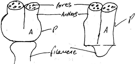
(b) Carefully remove one stamen from specimen M. Examine it using a hand lens. Draw and label it.
Conditions
P – Filament shorter than anther; ¼ of anther = filament.
A – All parts to be drawn; continuous lines.
(c) Remove another stamen from specimen M. Cut the anther transversely into two equal parts. Tap the pollen grains from the lower half onto a microscope slide. Add a drop of iodine. Place a cover slip and press on the cover slip gently to spread out the pollen grains. Observe the pollen grains under medium power.
 Draw one pollen grain.
State the magnification.
(d) Remove an anther from specimen N. Place it on a microscope slide. Add a drop of iodine. Cover with a cover slip. Press gently on the cover slip to spread out the pollen grains. Observe the pollen grains under medium power.
Draw one pollen grain.

State the magnification X 100.
(e) State two observable differences between the corolla of specimen N and M.
M – Smooth and small/smaller.
N – Rough/spiked and larger/larger.
(f) State four observable differences between the corolla of specimen M and N.
M – Petals fused – gamopetalous.
N – Free petals/overlapping corolla – polypetalous.
M – Small corolla.
N – Corolla large/broad.
M – Petals pointed tips.
N – Petals rounded tips.
M – Nectar guides not easily seen.
N – Nectar guides noticed.
2. Confidential requirement: Solution L – Diastase/amylase
You are provided with a solution labelled L, starch solution, and sodium chloride in two different concentrations 0.1% and 1.4%. Place 3 ml of starch solution in test tubes labelled 1, 2, and 3. Add 3 drops of 0.1% sodium chloride to the test tube labelled 3.
Add 3 ml of solution L to each test tube labelled 2 and 3.
- Place a drop of the contents from each test tube 1, 2, and 3 on a white tile. To each drop add iodine solution. Record your results in the table below.
| Test tube | Observation at start of experiment | Observation at end of experiment |
|---|---|---|
| Starch 1 | Blue – black Blue/black/dark blue | Blue-black/blue/black/dark blue |
| Starch + 0.1% NaCl + L 2 | Blue black as in TI | Retained the colour of iodine/yellow/brown/reddish/orange. Acc. Traces of blue Rej. Red |
| Starch + 1.4% NaCl + L 3 | Blue black as in TI | Retained iodine colour as in T2 |
- Place the test tube in a water bath maintained at 370C. Allow to stand for 30 minutes. Place a drop of the contents from each test on a white tile. To each drop add iodine solution. Record your observations in the table.
- Add equal amounts of Benedict’s Solution in test tubes labelled 2 and 3 and boil. Record your observations.
Test tube 2
Changed to green/yellow.
Test tube 3
Colour changed to orange/brown/red/reddish/brick red.
- Why was the test tube labeled 1 included in the experiment?
Control experiment.
- Account for the results in test tubes 1, 2, and 3 at the end of the experiment.
- Starch converted/hydrated/digested/broken down into sugars/reducing/glucose and maltose in test tubes 2 and 3.
- Starch was not converted into reducing sugars in test tube 1 due to lack of NaCl and enzyme (solution L).
- More reducing sugar in test tube 3 than in test tube 2 due to high concentration of NaCl in test tube 3.
- NaCl accelerates digestion/hydrolysis of starch.
- Suggest the identity of solution L.
Enzyme/diastase/amylase/ptyalin.
- Why were the test tubes placed in a water bath maintained at 370C? Provide optimum temperature/best temperature for enzyme activity (ideal/most suitable).
3. Confidential requirements: Specimen R – Housefly, Specimen S – Bee.
You are provided with specimens labeled R and S. Examine them.
- (i) Name the phylum and the class to which the specimens belong.
Phylum: Arthropoda
Class: Insecta
(ii) State two distinguishing features found in the members of:
Phylum: Presence of exoskeleton.
Joined appendages/limbs.
Class: 3 pairs of legs/six legs.
3 body parts: head, thorax, abdomen.
(b) State two differences between the wings of specimen R and S.
S: 2 pairs; absence of halteres/hind wings.
R: 1 pair of wings; has halteres/hind wings; modified wing.

Draw and label them.

(d) Draw and label the front view of the head of specimen S.


